 IMAGING FINDINGS:
Mammography shows an oval, hyperdense circumscribed mass with a few punctate calcifications. Breast ultrasound shows an intraductal mass with vascular flow. WHY IT MATTERS:
- Papilloma with atypia - Papillary DCIS - Papillary Carcinoma 3. May be solitary or multiple. If > 5 masses, can be considered papillomatosis. FURTHER READING: Breast Imaging Case of the Day (RadioGraphics, 1998)
0 Comments
 IMAGING FINDINGS:
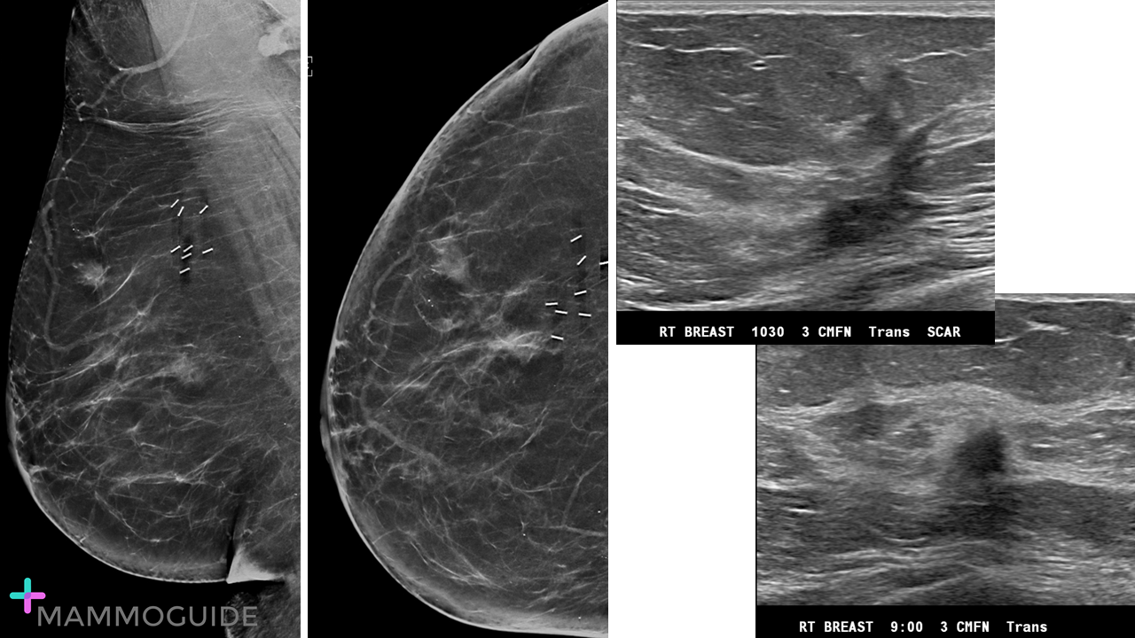
Mammography shows an irregular isodense mass adjacent to the prior lumpectomy scar. Ultrasound evaluation of the right breast shows surgical scarring at the 10:30 position an an irregular hypoechoic mass at the 9:00 position. WHY IT MATTERS:
b. Increasing density at the lumpectomy site c. New or increasing calcifications at the lumpectomy site FURTHER READING: The Postconservation Breast: Part 2, Imaging Findings of Tumor Recurrence and Other Long-Term Sequelae (AJR, 2012) 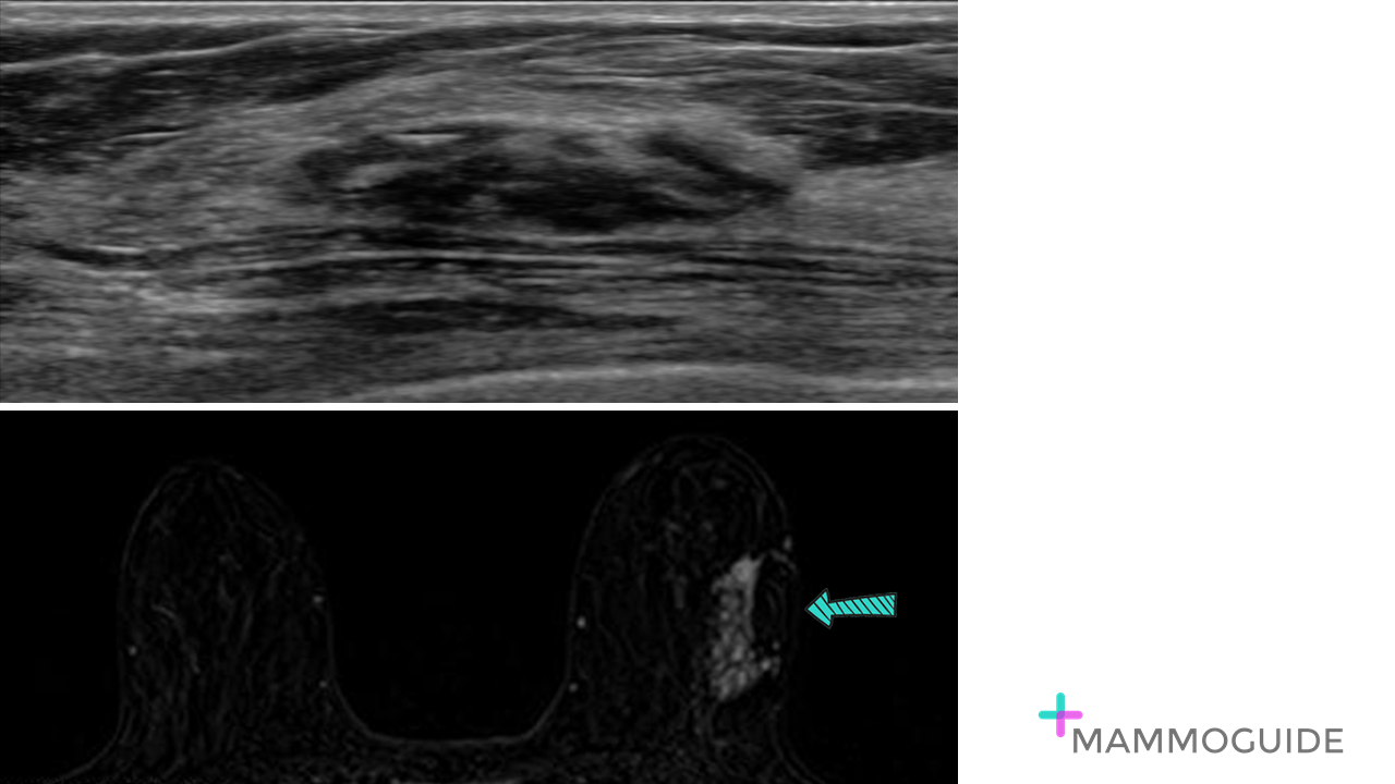 IMAGING FINDINGS:
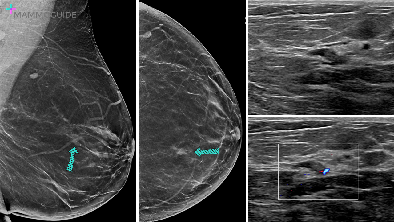
Breast ultrasound shows an oval heterogenous mixed echogenicity circumscribed mass that could easily be mistaken for normal fibroglandular tissue. Subtraction images on breast MRI demonstrate regional nonmass enhancement along the upper outer left breast. WHY IT MATTERS:
FURTHER READING: Pseudoangiomatous Stromal Hyperplasia: Imaging Findings With Pathologic and Clinical Correlation (AJR, 2010) 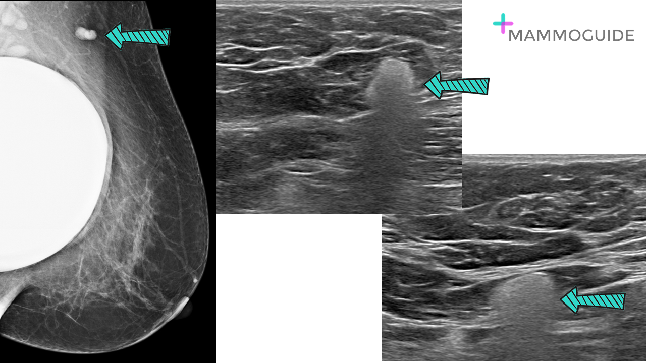 IMAGING FINDINGS:
Abnormal lymph node with hyperdense appearance on mammography. A sub-pectoral silicone implant is also noted. On ultrasound, there is a hazy, "snow storm" like appearance of increased echogenicity within a lymph node consistent with free (extra-capsular) silicone. WHY IT MATTERS:
FURTHER READING: Multimodality Imaging-based Evaluation of Single-Lumen Silicone Breast Implants for Rupture (RadioGraphics, 2017) 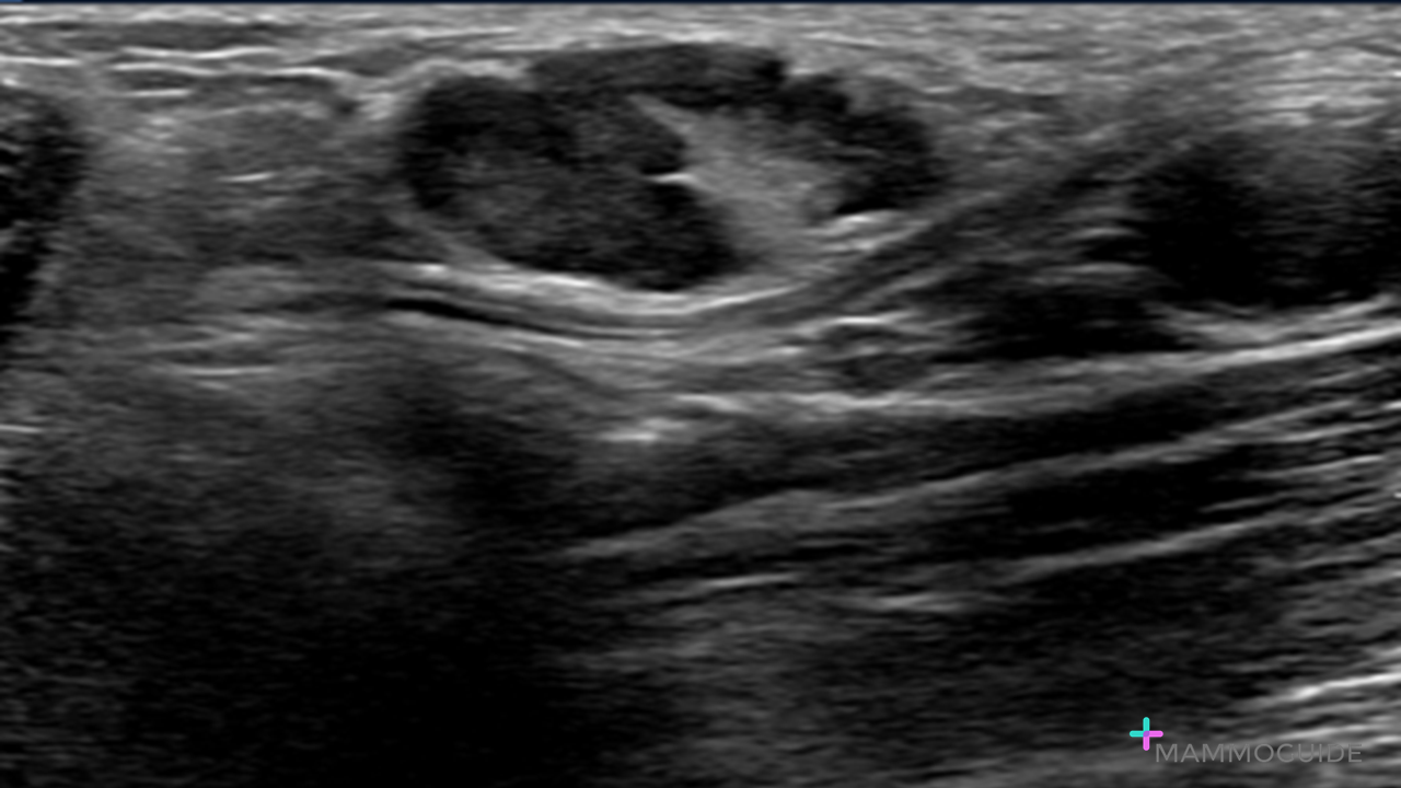 IMAGING FINDINGS:
Abnormal enlarged axillary lymph node with an irregular asymmetrically thickened cortex. WHY IT MATTERS:
- focal cortical bulge or eccentric thickening - irregular margins or encroachment of the fatty hilum - loss of the normal fatty hilum For axillary lymph nodes on ultrasound, the overall size (length or width) of the node is not the most important factor. It is the cortical thickness that matters. FURTHER READING: US Evaluation of Axillary Lymph Nodes (RadioGraphics, 2014) |
Quick CasesA picture is worth a thousand words. High-yield examples of essential breast imaging knowledge. 
Categories
All
|
 RSS Feed