 IMAGING FINDINGS:
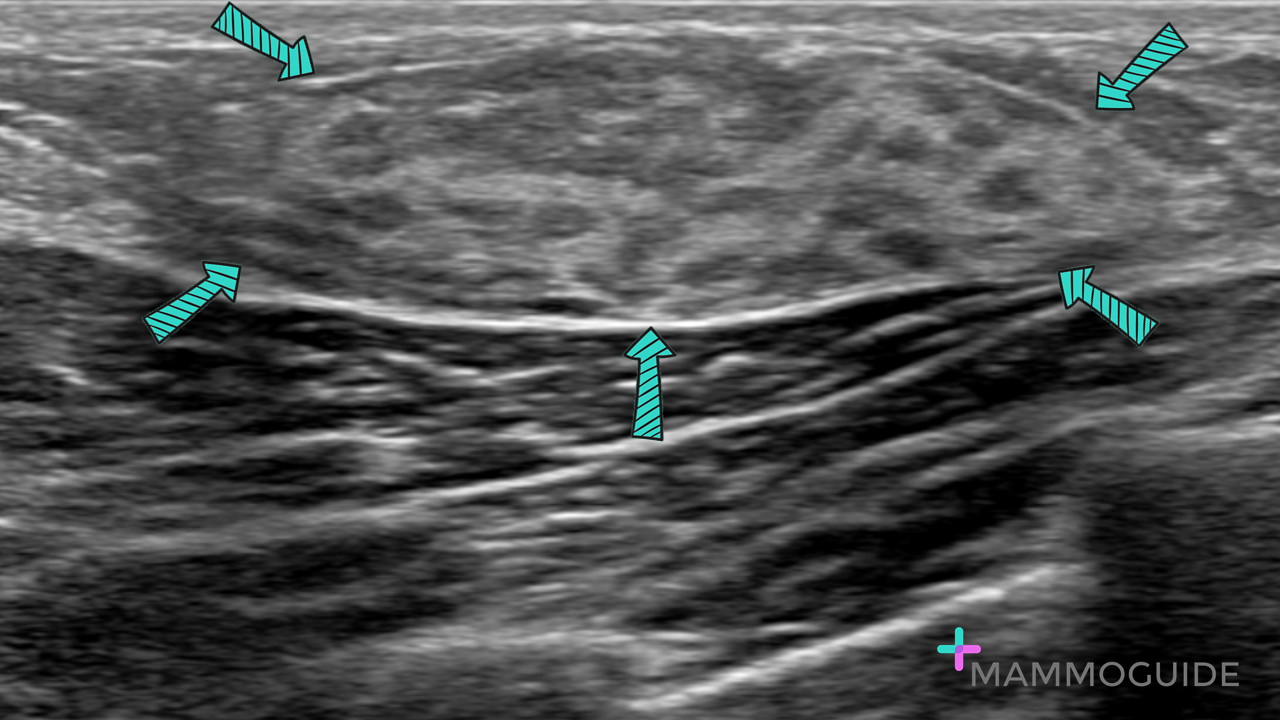
Sonographic image of a palpable area of concern in the left axilla demonstrates normal dense fibroglandular tissue consistent with prominent accessory breast tissue. WHY IT MATTERS:
FURTHER READING: The ABCs of Accessory Breast Tissue: Basic Information Every Radiologist Should Know (AJR 2014)
0 Comments
 IMAGING FINDINGS:
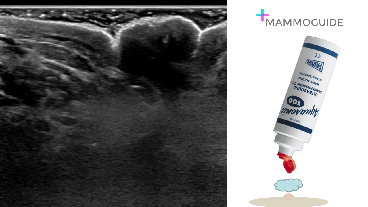
Sonographic image of the nipple and subareolar region shows no abnormality. Notice the large amount of ultrasound gel used to adequately image the subareolar region. WHY IT MATTERS:
FURTHER READING: Nipple-Areolar Complex: Normal Anatomy and Benign and Malignant Processes (RadioGraphics 2009) 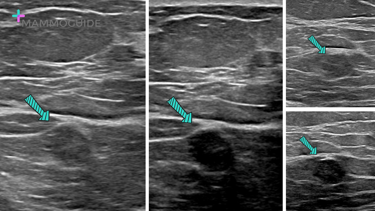 IMAGING FINDINGS:
Sonographic image of the right breast with and without the use of harmonics demonstrate an oval isoechoic mass with irregular margins. The mass is much easier to see with harmonics on. WHY IT MATTERS:
FURTHER READING: A Primer on the Physical Principles of Tissue Harmonic Imaging (RadioGraphics 2015) 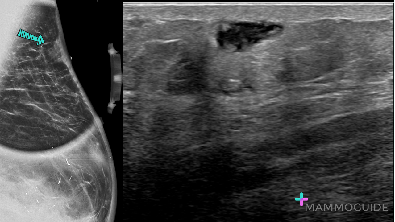 IMAGING FINDINGS:
Tangential spot MLO view of a palpable lump in the left breast shows a superficial mass with ill-defined margins. Ultrasound evaluation of the palpable area of concern demonstrates an irregular mixed echogencity superficial mass. WHY IT MATTERS:
Radiation-Induced Sarcoma of the Breast: A Systematic Review (The Oncologist, 2012) 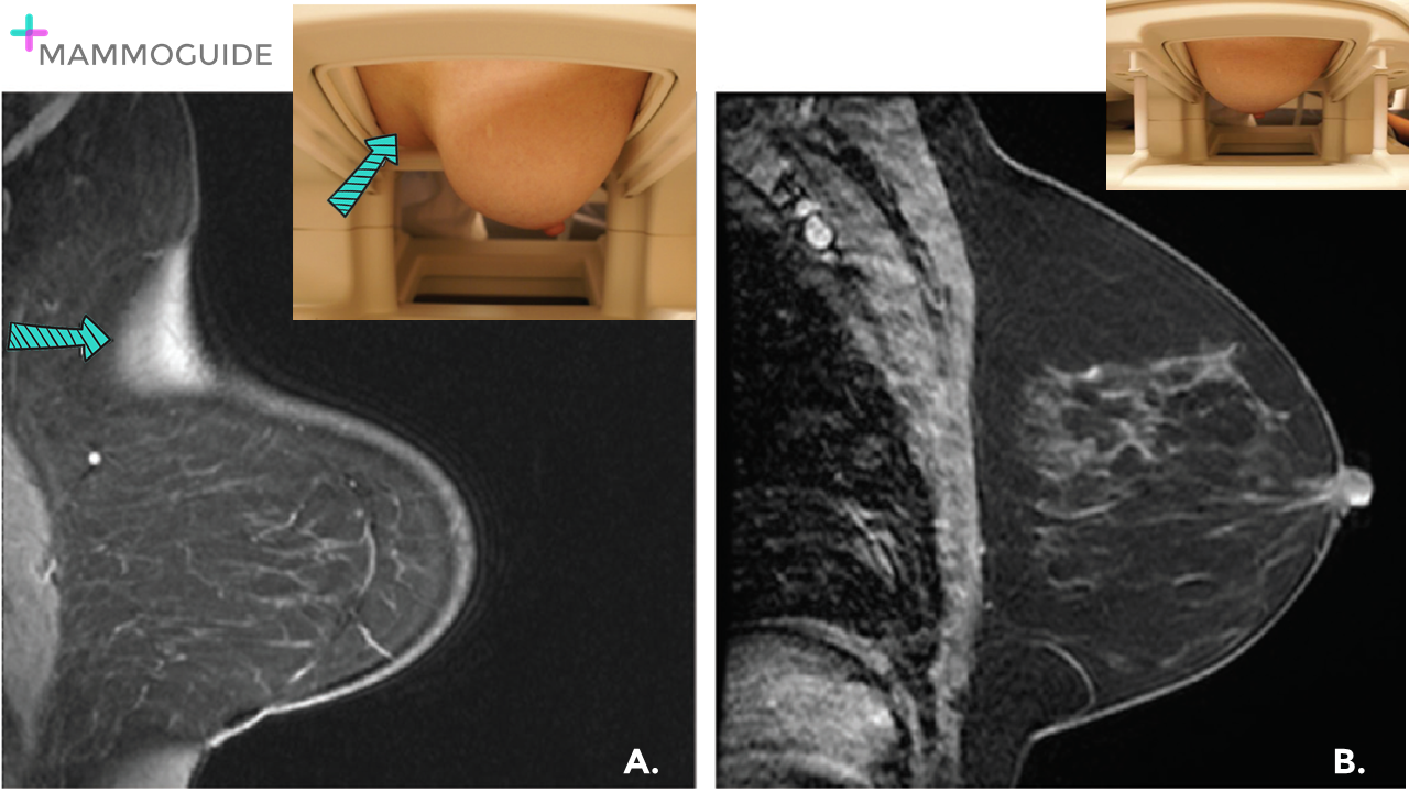 IMAGING FINDINGS:
Sagittal fat saturated T1-weighted image (A.) demonstrates poor fat saturation due to coil artifact in the superior left breast. The breast is positioned poorly with too much of the abdomen included in the coil, forcing the superior breast to touch the coil. After the patient is re-positioned with the breast centered in the coil (B.), there is no longer coil artifact. WHY IT MATTERS:
FURTHER READING: Breast MR Imaging Artifacts: How to Recognize and Fix Them (RadioGraphics 2007)  IMAGING FINDINGS:
Axial and Sagittal post-contrast images demonstrate linear non-mass enhancement in the central right breast. WHY IT MATTERS:
FURTHER READING: Nonmass Enhancement on Breast MRI: Review of Patterns With Radiologic-Pathologic Correlation and Discussion of Management (AJR 2015) 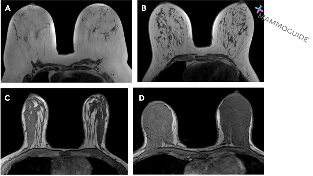 IMAGING FINDINGS:
Four selected axial non-fat saturated T1-weighted images, from four different patients, reveal the differences in fibroglandular tissue.
WHY IT MATTERS:
FURTHER READING: ACR BI-RADS® Atlas Fifth Edition 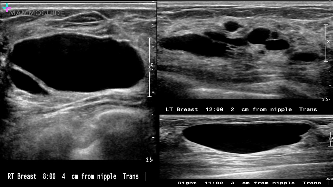 IMAGING FINDINGS:
Selected ultrasound images show multiple simple cysts scattered throughout the breasts bilaterally. There is a background of dense fibroglandular tissue. WHY IT MATTERS:
FURTHER READING: Complex Cystic Breast Masses: Diagnostic Approach and ImagingPathologic Correlation (RadioGraphics 2007)  IMAGING FINDINGS:
Standard Craniocaudal (CC) and Mediolateral Oblique (MLO) views of the left breast (A and B) demonstrate heterogeneously dense breast tissue with no definite abnormality. Selected Craniocaudal tomosynthesis image (C.) shows an irregular spiculated mass in the lateral left breast. WHY IT MATTERS:
FURTHER READING: Outcome of Architectural Distortion Detected Only at Breast Tomosynthesis versus 2D Mammography (Radiology 2018) 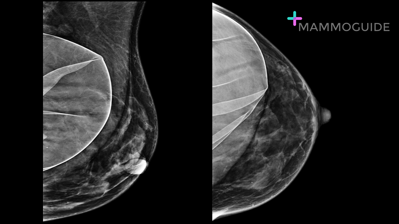 IMAGING FINDINGS:
Craniocaudal and mediolateral oblique views of the left breast demonstrate a subpectoral saline breast implant. The implant is ruptured. WHY IT MATTERS:
FURTHER READING: The Augmented Breast: A Pictorial Review of the Abnormal and Unusual (AJR 2011) |
Quick CasesA picture is worth a thousand words. High-yield examples of essential breast imaging knowledge. 
Categories
All
|
 RSS Feed