Finding a subtle, subcentimeter area of architectural distortion on a screening mammogram can feel really good.
But oftentimes, the sense of accomplishment gives way to a sense of dread as you realize you now have to figure out how to sample this lesion.
The vast majority of breast biopsies performed today are percutaneous core biopsies. These quick, safe and accurate procedures have nearly completely replaced open surgical biopsies or Fine-Needle Aspirations (FNA). While the procedures are relatively simple, there are a few places where you can get tripped up if you are not paying attention.
Biopsy Needle Options
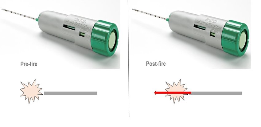
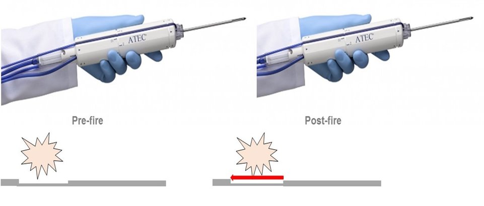
Breast core biopsy needles come in a variety of sizes. Needle size is measured in gauge (thickness) and length. In general, the most common length of biopsy needles are either 10cm or 16cm. The more important measurement is the thickness or gauge of the needle.
The most common needle gauges used in the breast vary depending on the type of biopsy. Ultrasound core biopsies typically use a 14g or 12g needle. Stereotactic core biopsies typically use larger needles such as 7g, 9g or 12g. MRI biopsies usually use 9g or 12g needles as well.
Type of Breast Biopsy Guidance
Core biopsies of the breast can be performed using ultrasound, mammographic, or MRI guidance. In general, the modality on which you can see the lesion best is the optimal way to perform the procedure. That being said, ultrasound core biopsies are the quickest and most cost effective type of breast intervention and should be your first choice if at all possible.
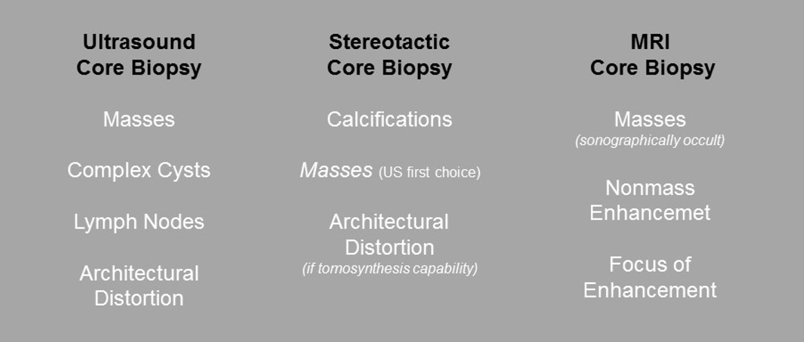
Figure 1. Best Way to Biopsy Specific Breast Lesions
There are two main types of core biopsy devices. These include spring loaded devices and vacuum assisted biopsy devices. Spring loaded devices have a needle that fires through the lesion to acquire the core sample. They have the advantage of being cheaper, lighter and easier to maneuver, and easier to set up and use. They are ideal for most ultrasound core biopsies. However, they should never be used for stereotactic or MRI guided biopsies.
Spring Loaded Breast Biopsy Device
Some of the most common spring loaded breast biopsy devices used in the U.S. include: Bard Monopty 14g or 12g Bard Max Core 14g Hologic Sertera 14g Hologic Tru-Core 14g Bard Mission 14g Achieve 14g
Vacuum assisted breast biopsy devices have a needle with an aperture that is placed under or within the lesion of interest. A vacuum sucks tissue into the aperture and then the aperture closes to complete tissue acquisition. They tend to be larger needles and because of the vacuum usually obtain larger size tissue samples. Most of the vacuum assisted devices have a separate console that is hooked to the biopsy device with suction tubing. They are ideally used for all stereotactic and MRI guided core biopsies. Occasionally, a vacuum assisted device can also be useful when performing an ultrasound core biopsy of complex cysts, papillary lesions or small areas of architectural distortion.
Vacuum Assisted Breast Biopsy Device
Some of the most common vacuum assisted biopsy devices used in the U.S. include: Suros ATEC Breast Biopsy System 9g or 12g Eviva Breast Biopsy System 9g or 12g EnCor Enspire Breast Biopsy System 7g, 10g, or 12g Mammotome Breast Biopsy System 8g or 10g
There are also a number of "pseudo-vacuum" assisted devices. These are self-contained (not hooked up to a vacuum console) and easy to use like the spring loaded devices, yet provide slightly better volume cores due to a small amount of vacuum suction. They work great for ultrasound core biopsies. Because they are slightly more expensive, they have fallen out of favor lately due to changes in breast biopsy reimbursement.
Some of the most common "pseudo-vacuum" assisted handheld devices used in the U.S. include: Hologic Celero 12g Bard Vacora 10g or 14g Bard Finesse Ultra 10g or 14g
Clip Placement
The final step of any breast biopsy is placing a biopsy clip. These small markers (generally made of titanium or other alloys) are placed at the site of biopsy. No matter the outcome of the biopsy, benign or malignant, they are very useful. They are used to mark the area in case further intervention (i.e. surgical excision) is required. If the biopsy is benign and no further intervention is required, the markers simply stay put and show up on future imaging to denote that the area has already been sampled. This avoids repeat biopsy of the same area.
Want to learn more? Check out our signature training course.