|
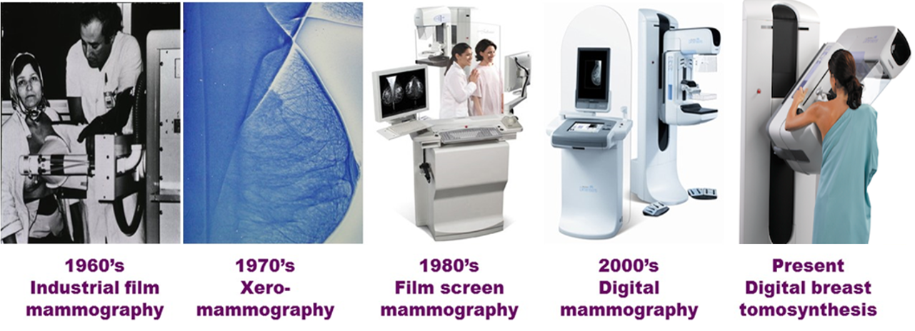
Digital Breast Tomosynthesis, sometimes referred to as 3D Mammography, is an exciting new technique for performing breast imaging.
It is quickly replacing traditional mammography as the go-to screening & diagnostic exam for evaluating the breast. In the sections that follow we will go through several basic concepts regarding tomosynthesis. We will also cover the many uses, benefits and drawbacks of implementing this exciting new technology. |

|
|
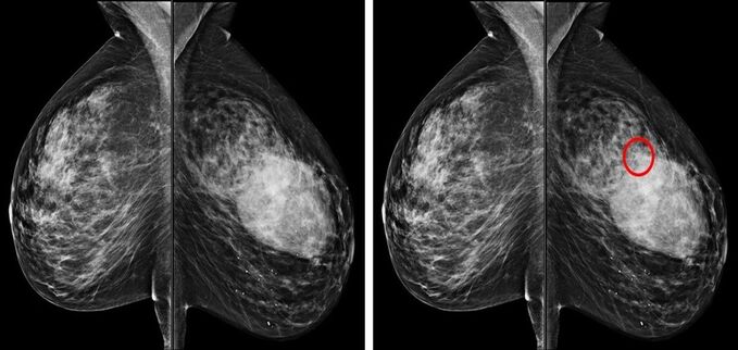
Overlap and Superimposition False Negative Findings = Missed Cancers False Positive Findings = Unnecessary Recalls |
|
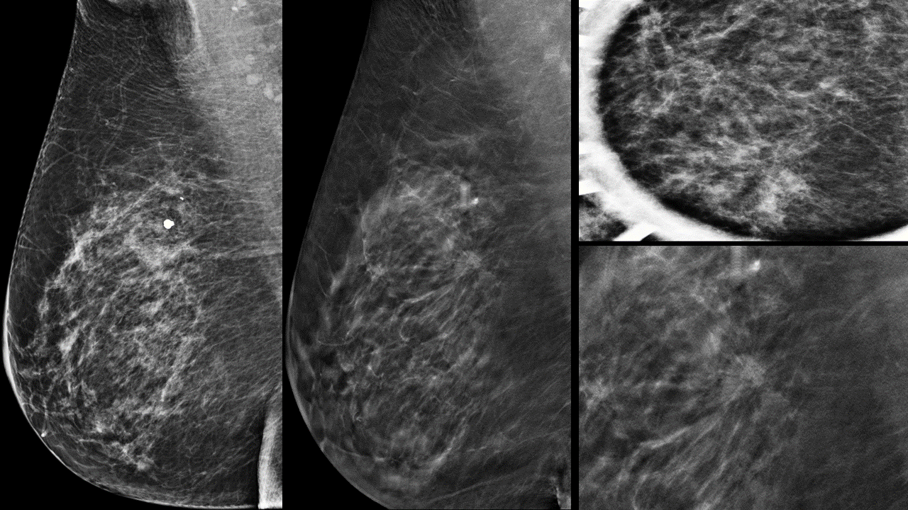
While trying to find a small mass in a busy pattern of dense breast tissue can seem like searching for a needle in a haystack, if you remove the overlapping structures the cancer becomes readily apparent. This is the observation made by a team at Mass General Hospital (MGH) in Boston led by Dr. Daniel Kopans. They realized that when performing mammography on specimens from patients who underwent lumpectomies, it was very easy to see the mass in great detail. This was because the overlapping structures were removed.
|
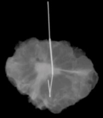
Radiograph of a lumpectomy specimen
|
|
|
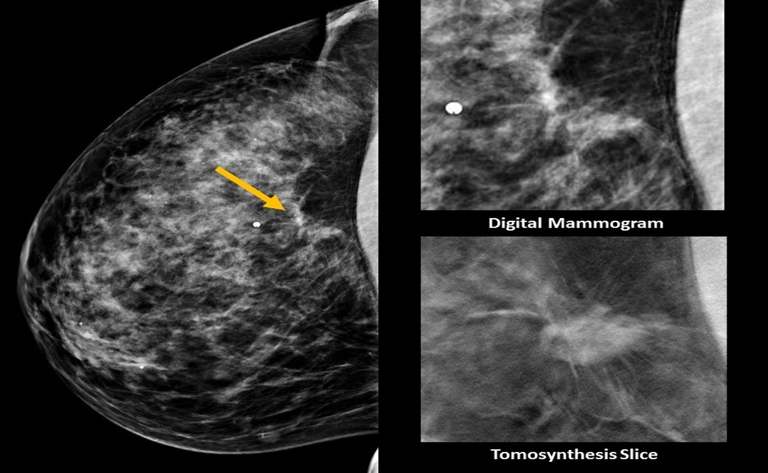
Imagine a book that you want to read, but imagine that it is unique in that it has clear pages. Now try and read it by looking only at the first page. You will see thousands of words, but they will look all jumbled because they all are overlapping each other. It is tough to make sense of anything. This is what reading a standard mammogram is like! Interpreting a tomosynthesis exam is like taking that same book, but instead of only looking at the first page, you flip through it page by page. Doing this makes reading the book so much easier. |
|
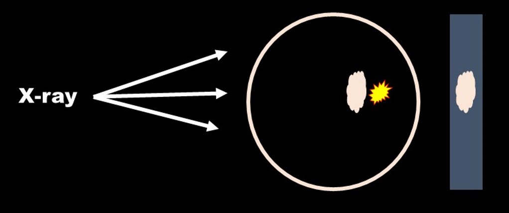
For a tomosynthesis examination, the breast is positioned and placed in compression. This is essentially the same as for a traditional mammogram. The difference is that the x-ray tube rotates through a 15 degree arc and acquires 15 projection images. These projection images are then reconstructed into a series of slices through the breast. A standard tomosynthesis view will require anywhere from 40 - 100+ slices to cover the entire breast. The number of slices varies depending on the size of the breast. Thicker breasts = more tomo slices.
The last step of a tomosynthesis exam is that while the patient is still in compression a traditional 2D image is then acquired. Next, the compression is released and the patient re-positioned for the next view. The tomosynthesis images are to be scrolled through, similar to a CT or MRI exam. They also can be viewed as a cine loop. The interpreting radiologist therefore has the standard 4 views (CC + MLO) plus 4 tomosynthesis cine loops to scroll through. |
|
|
Tomosynthesis is a very unique imaging exam in that it increases the sensitivity and specificity of mammography. What this means is that it increases cancer detection but also decreases false positives (unnecessary extra views). This is rare.
Adding breast ultrasound or breast MRI to a screening regimen will find more cancers but with the trade-off of more false positives. You will also find and have to biopsy more benign lesions. Tomosynthesis is different. Most screening mammography recalls and extra views are for normal overlapping tissue. But, when you screen with tomosynthesis, the frequency of these unnecessary (false positive) exams, decreases significantly. Multiple studies have consistently shown increased cancer detection and decreased false positives when tomosynthesis is implemented. The exact percent change varies based on the study, but the numbers are huge. |

|