Five Ways Not to Fail |

|
|
Don’t fail.
Whether it is at the ABR core exam, in the reading room or in the courtroom for a malpractice suit, following a few simple breast imaging rules will keep you out of trouble. These 5 rules may seem like no-brainers and are often glossed over, however if you fail to obey these guidelines even once, you are putting yourself and your patients at risk. |
|
|
Rule #1
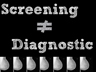
The most basic rule, but it must never be violated. A screening exam is just that, screening. The patient may not have any palpable areas, nipple discharge, focal pain or any other issue. Screening exam = only CC and MLO views (occasionally a XCCL may be required to visualize all the tissue). |
|
|
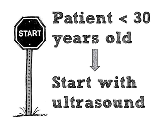
Rule #2
A standard breast imaging diagnostic exam begins with mammography. However, the key exception is a female patient under age 30 (some facilities use age 28 instead of 30). Ultrasound is the first step in this young patient population. |
|
|
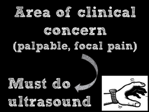
Rule #3
Any patient who presents with an area of clinical concern, must be evaluated with ultrasound in addition to diagnostic mammography. If a patient feels a lump or six lumps, you are obligated to put the ultrasound probe on each site. Breast thickening, a lump felt by another doctor and focal pain (able to point to with one finger) also require sonographic evaluation. Failure to perform an ultrasound goes against the current standard of care. |
|
|
Rule #4
Breast imaging is one of the most regulated, orderly specialties in all of medicine. You should have a standard protocol for evaluating every possible scenario. A palpable lump should be imaged the same every time. The same for calcifications, architectural distortion or any other mammographic abnormality. |

|
|
Rule #5
Make sure all your terminology is consistent. The American College of Radiology has laid out very specific descriptors that are to be used to describe each finding in the breast (whether on a mammogram, ultrasound or MRI). If you use a word like “irregular” or “spiculated,” you better make sure you are giving a BIRADS 4 or 5 and recommending a biopsy. |

|