 IMAGING FINDINGS:
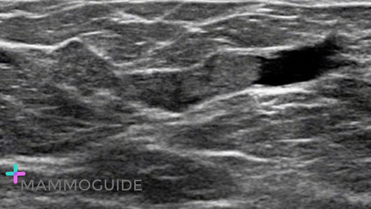
Sonographic image demonstrates a dilated duct with an intraductal mass or filling defect in the subareolar left breast. WHY IT MATTERS:
FURTHER READING: Kim W.H, Chang J.M., et al. Intraductal Mass on Breast Ultrasound: Final Outcomes and Predictors of Malignancy. AJR 2013; 200:932–937
0 Comments
 IMAGING FINDINGS:
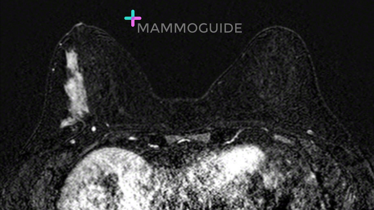
Axial subtraction image demonstrates homogenous segmental non-mass enhancement in the lateral right breast. WHY IT MATTERS:
FURTHER READING: ACR BI-RADS Atlas Fifth Edition 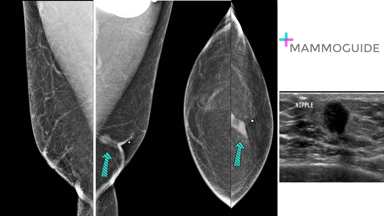 IMAGING FINDINGS:
Mediolateral oblique (MLO) and craniocaudal (CC) views of both breasts demonstrate an irregular mass in the subareolar left breast. There is associated nipple retraction. Corresponding ultrasound image shows a round mass with subtle spiculated margins. WHY IT MATTERS:
FURTHER READING: Male Breast Cancer in the Age of Genetic Testing: An Opportunity for Early Detection, Tailored Therapy, and Surveillance (RadioGraphics 2018) 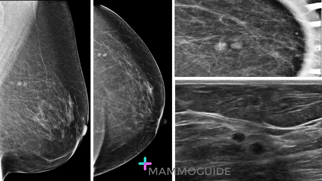 IMAGING FINDINGS:
Mediolateral oblique (MLO) and craniocaudal (CC) views of the left breast demonstrate two round circumscribed masses in the upper outer quadrant posteriorly. Spot compression view confirms the finding. Ultrasound evaluation demonstrates two adjacent round hypoechoic masses. They do not have the characteristics of a lymph node. Biopsy revealed multifocal invasive ductal carcinoma. WHY IT MATTERS:
Intramammary Lymph Nodes: Normal and Abnormal Multimodality Imaging Features (BJR 2019) 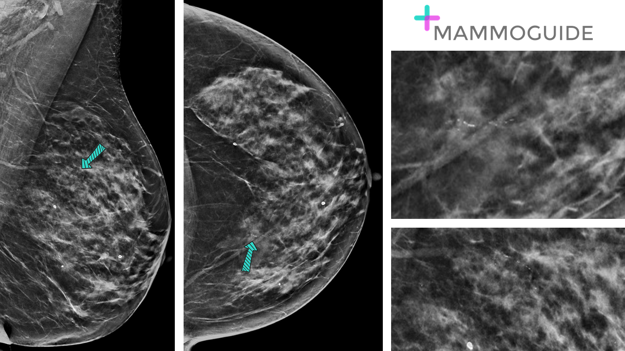 IMAGING FINDINGS:
Standard craniocaudal (CC) and mediolateral oblique (MLO) views demonstrate linear fine pleomorphic calcifications in the upper inner left breast. WHY IT MATTERS:
FURTHER READING: Radiologic-Pathologic Correlation of Ductal Carcinoma in Situ (RadioGraphics 2010) 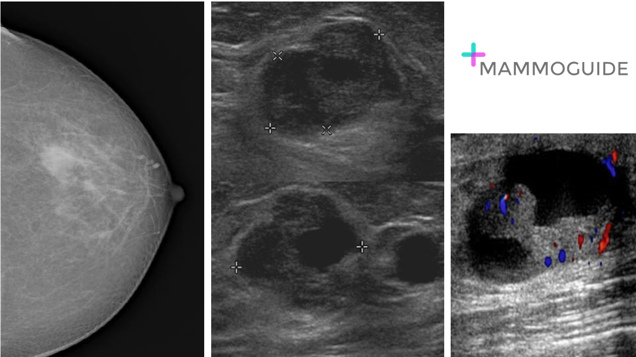 IMAGING FINDINGS:
Single craniocaudal mammographic view (CC) of the left breast demonstrates a mass in the lateral left breast. Selected sonographic images show a complex cyst. The complex cyst has anechoic cystic and hypoechoic solid components. There is vascular flow within the solid component. WHY IT MATTERS:
FURTHER READING: Complex Cystic Breast Masses: Diagnostic Approach and Imaging-Pathologic Correlation (RadioGraphics 2007) 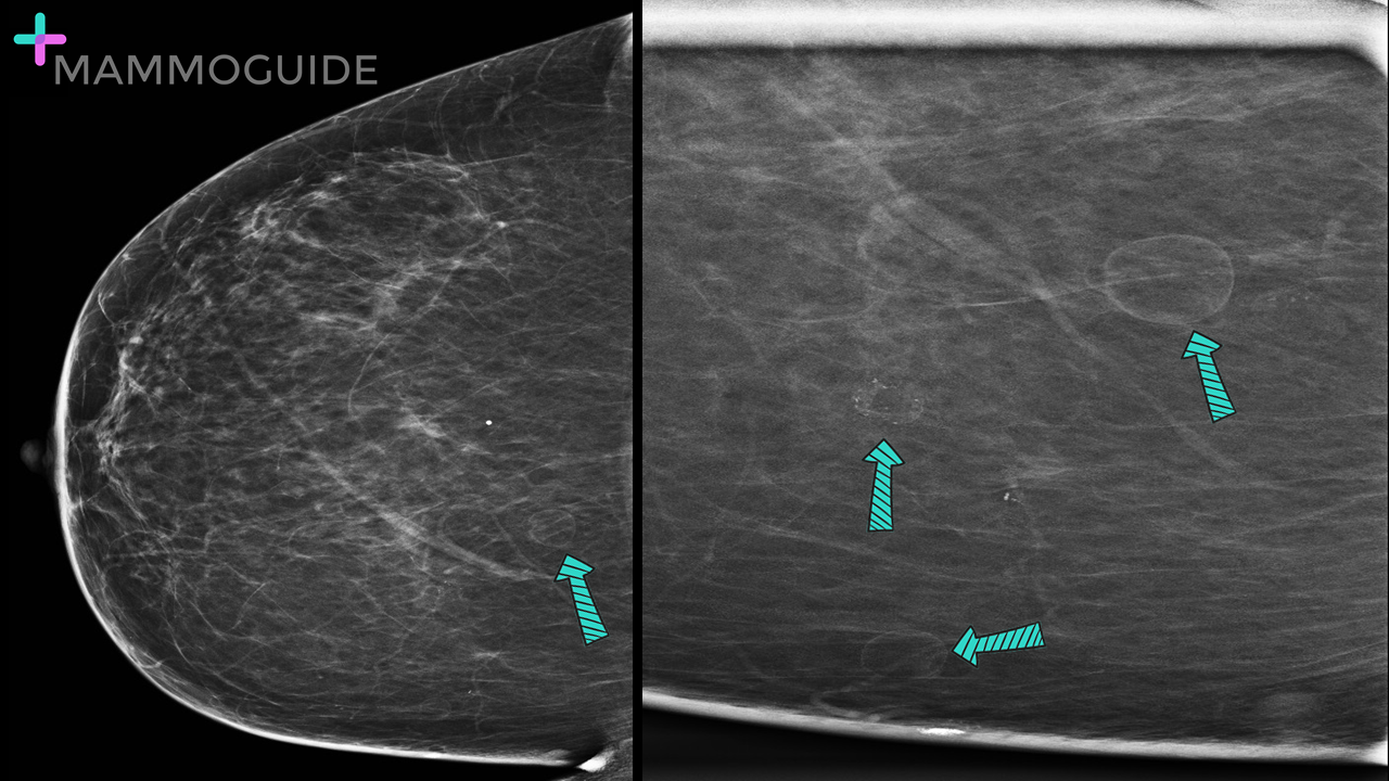 IMAGING FINDINGS:
Craniocaudal and quad magnification view of the right breast demonstrate multiple fat density circumscribed masses consistent with benign oil cysts. WHY IT MATTERS:
FURTHER READING: Cystic Masses of the Breast (AJR 2010) 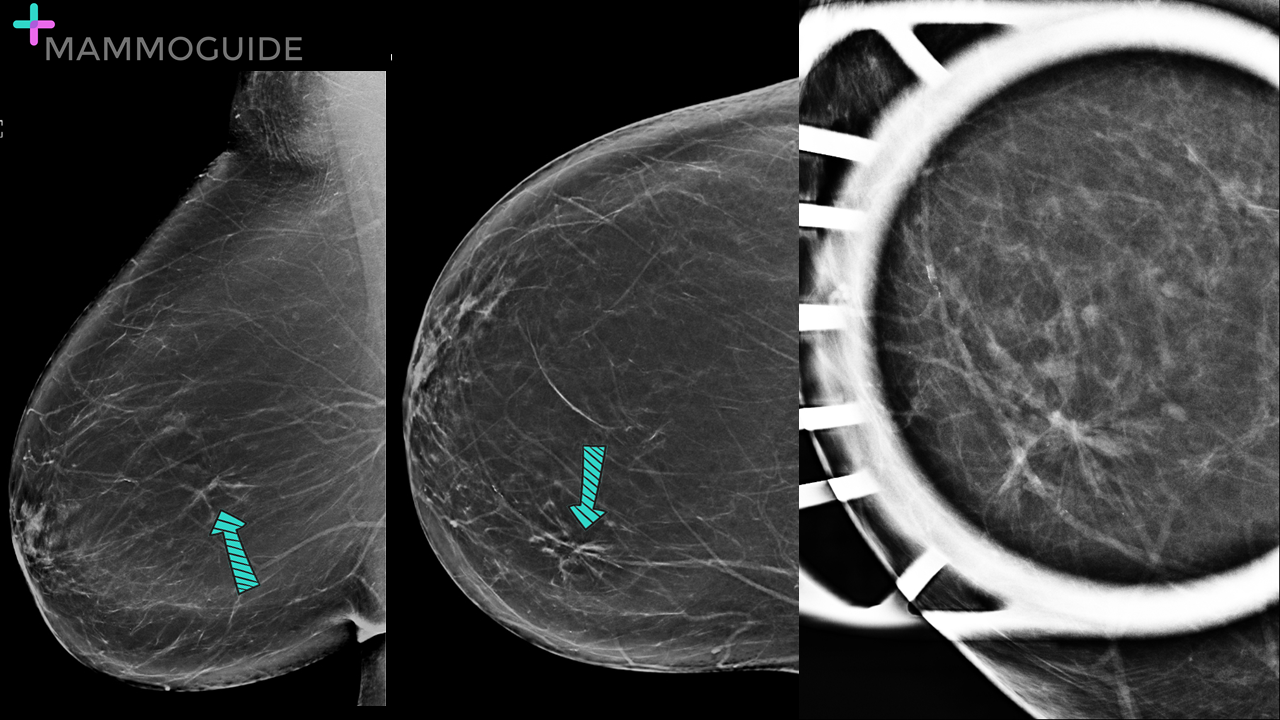 IMAGING FINDINGS:
Craniocaudal (CC) and Mediolateral Oblique (MLO) views of the right breast demonstrate an area of architectural distortion medially. Spot compression view confirms persistence of architectural distortion. WHY IT MATTERS:
FURTHER READING: Radial Scars/Complex Sclerosing Lesions of the Breast: Radiologic and Clinicopathologic Correlation (BMC Medical Imaging 2018) 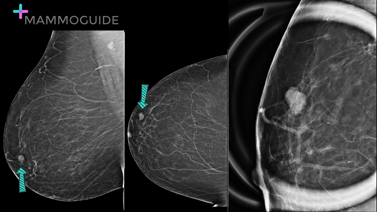 IMAGING FINDINGS:
Standard craniocaudal (CC) and mediolateral oblique views of the right breast demonstrate a subcentimeter mass in the lateral right breast. Spot magnification view of the high density mass shows suspicious microlobulated margins. WHY IT MATTERS:
FURTHER READING: ACR BI-RADS® Atlas Fifth Edition QUICK REFERENCE 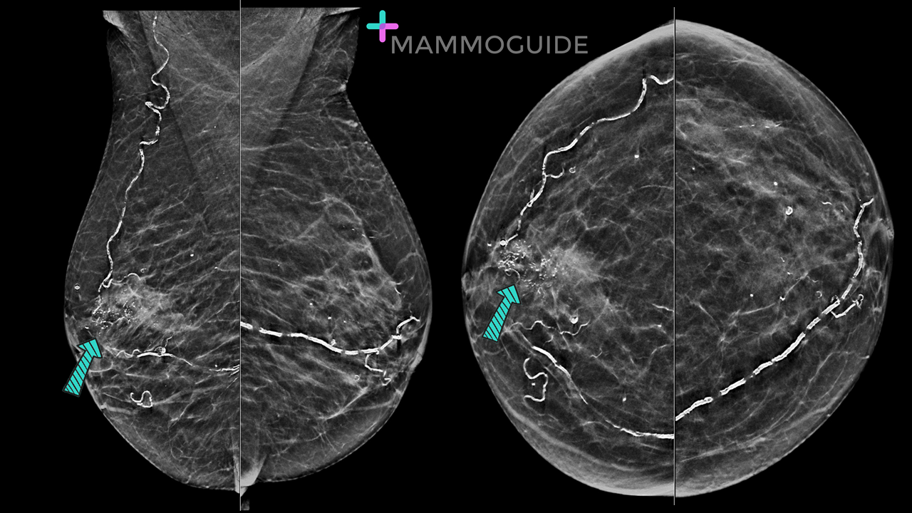 IMAGING FINDINGS:
Standard craniocaudal (CC) and mediolateral oblique views of both breasts demonstrate a grouping of fine pleomorphic calcifications in the subareolar right breast. WHY IT MATTERS:
ACR BI-RADS® Atlas Fifth Edition QUICK REFERENCE |
Quick CasesA picture is worth a thousand words. High-yield examples of essential breast imaging knowledge. 
Categories
All
|
 RSS Feed