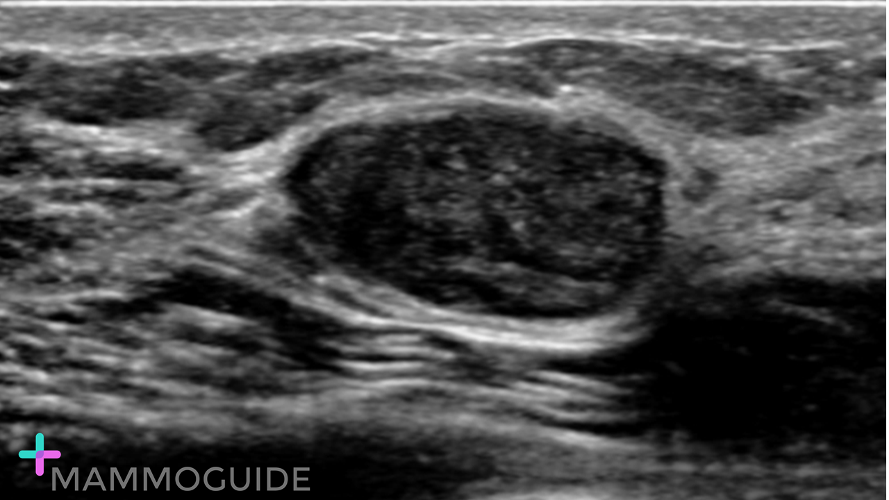
 IMAGING FINDINGS:
Single ultrasound image demonstrates an oval, circumscribed hypoechoic mass. The mass is wider than tall and does not demonstrate any suspicious features. WHY IT MATTERS:
Fibrous Lesions of the Breast: Imaging-Pathologic Correlation (RadioGraphics 2005)
0 Comments
Leave a Reply. |
Quick CasesA picture is worth a thousand words. High-yield examples of essential breast imaging knowledge. 
Categories
All
|
 RSS Feed