 IMAGING FINDINGS:
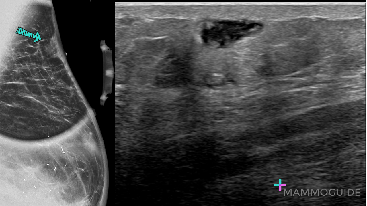
Tangential spot MLO view of a palpable lump in the left breast shows a superficial mass with ill-defined margins. Ultrasound evaluation of the palpable area of concern demonstrates an irregular mixed echogencity superficial mass. WHY IT MATTERS:
Radiation-Induced Sarcoma of the Breast: A Systematic Review (The Oncologist, 2012)
0 Comments
 IMAGING FINDINGS:
Standard Craniocaudal (CC) and Mediolateral Oblique (MLO) views of the left breast (A and B) demonstrate heterogeneously dense breast tissue with no definite abnormality. Selected Craniocaudal tomosynthesis image (C.) shows an irregular spiculated mass in the lateral left breast. WHY IT MATTERS:
FURTHER READING: Outcome of Architectural Distortion Detected Only at Breast Tomosynthesis versus 2D Mammography (Radiology 2018) 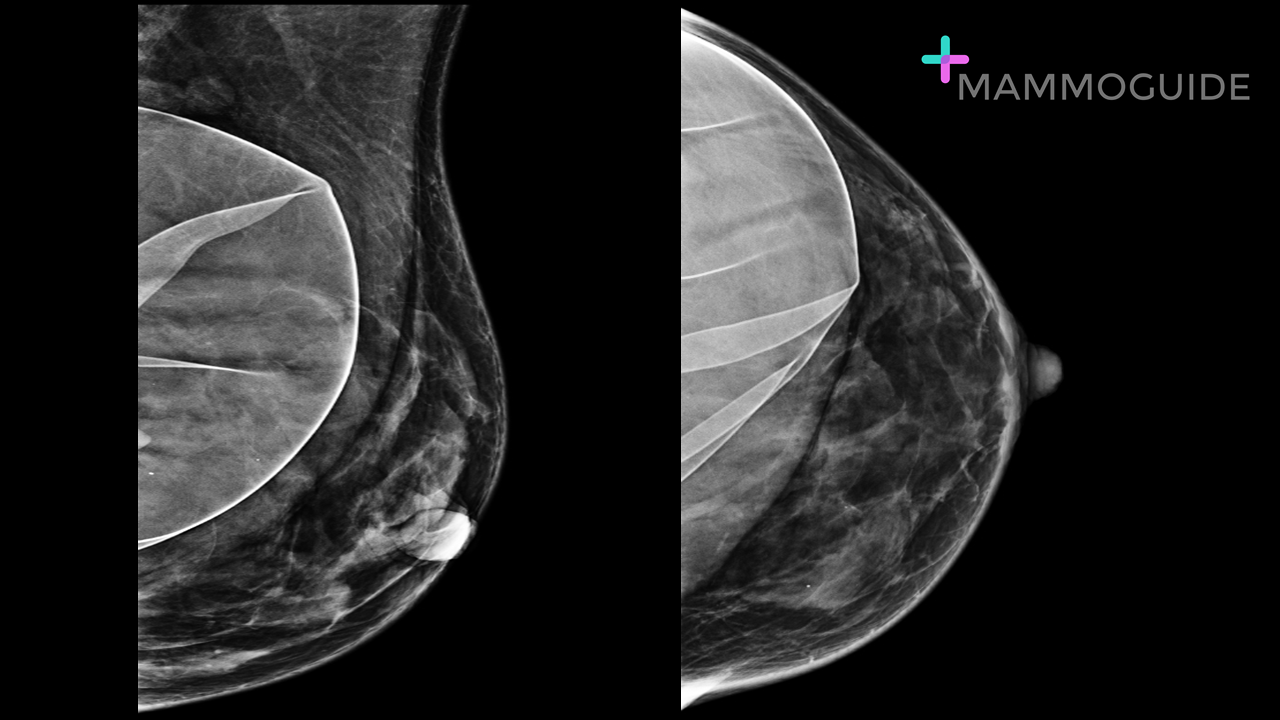 IMAGING FINDINGS:
Craniocaudal and mediolateral oblique views of the left breast demonstrate a subpectoral saline breast implant. The implant is ruptured. WHY IT MATTERS:
FURTHER READING: The Augmented Breast: A Pictorial Review of the Abnormal and Unusual (AJR 2011) 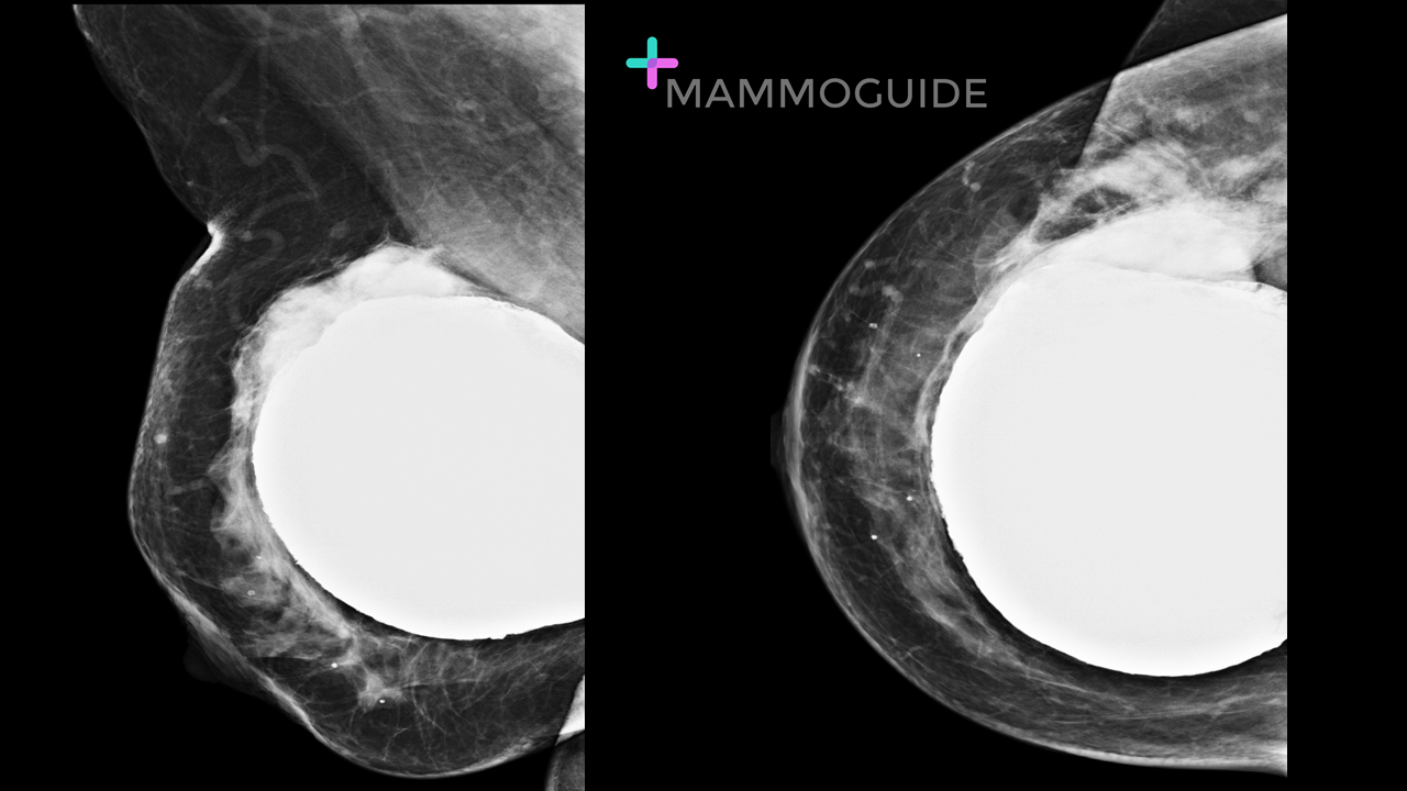 IMAGING FINDINGS:
Craniocaudal and mediolateral oblique views of the right breast demonstrate a subglandular silicone implant rupture. The implant is deformed with high density free silicone present along the upper outer margin. WHY IT MATTERS:
FURTHER READING: The Augmented Breast: A Pictorial Review of the Abnormal and Unusual (AJR 2011) 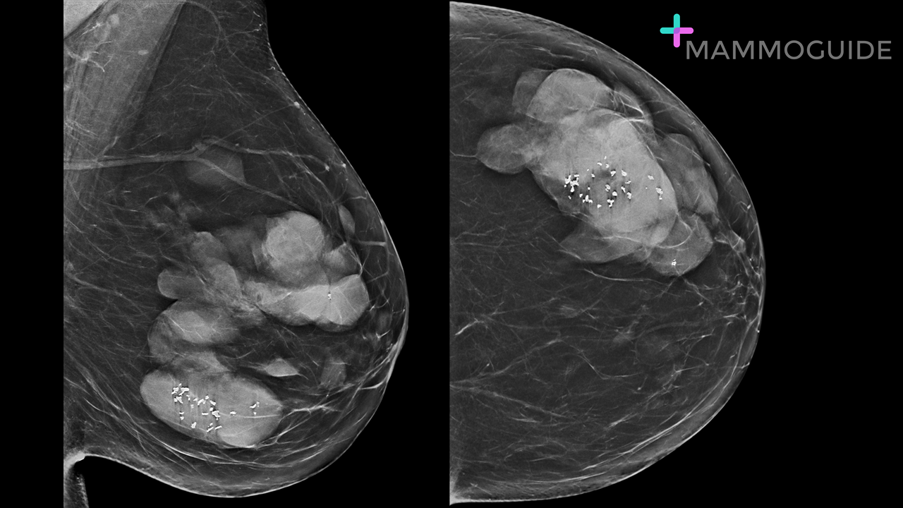 IMAGING FINDINGS:
Craniocaudal and mediolateral oblique views of the left breast show a large circumscribed mixed density mass occupying the upper outer quadrant. There are fat, calcifications and soft tissue density components to the mass. WHY IT MATTERS:
FURTHER READING: Unusual Breast Lesions: Radiologic-Pathologic Correlation (RadioGraphics 1999) 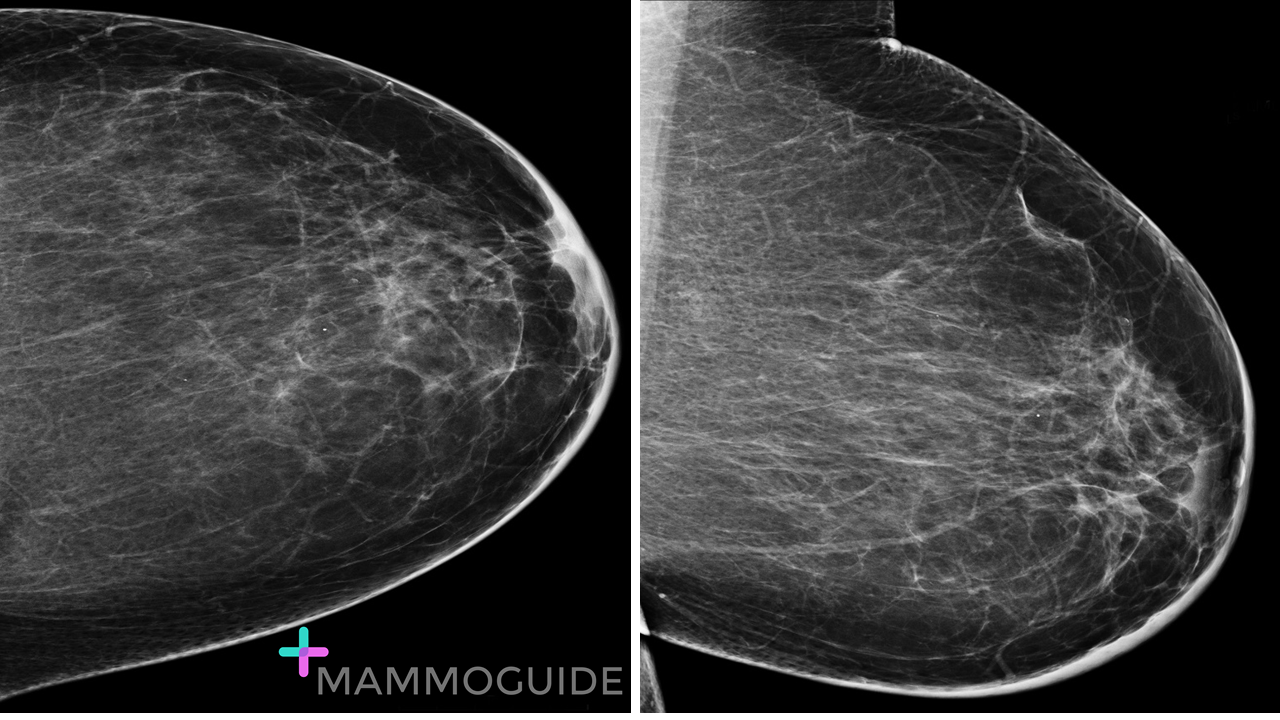 IMAGING FINDINGS:
Craniocaudal and mediolateral oblique views of the left breast demonstrate diffuse skin thickening, more prominent in the lower inner quadrant. No mass or suspicious calcifications are identified. WHY IT MATTERS:
What Radiologists Need to Know about Diagnosis and Treatment of Inflammatory Breast Cancer: A Multidisciplinary Approach (RadioGraphics 2013) 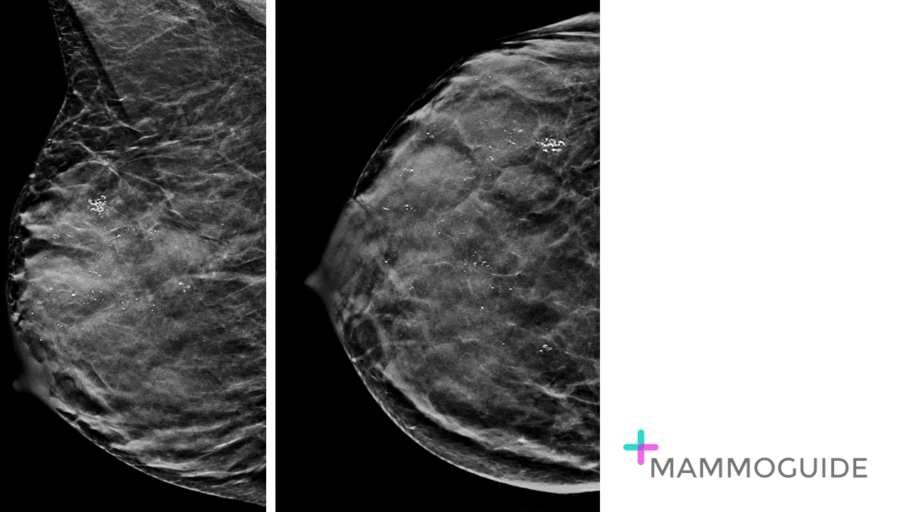 IMAGING FINDINGS:
Craniocaudal and mediolateral oblique views of the right breast demonstrate diffuse punctate calcifications throughout the breast. No masses or architectural distortion is identified. WHY IT MATTERS:
FURTHER READING: ACR BI-RADS Atlas Fifth Edition Quick Reference 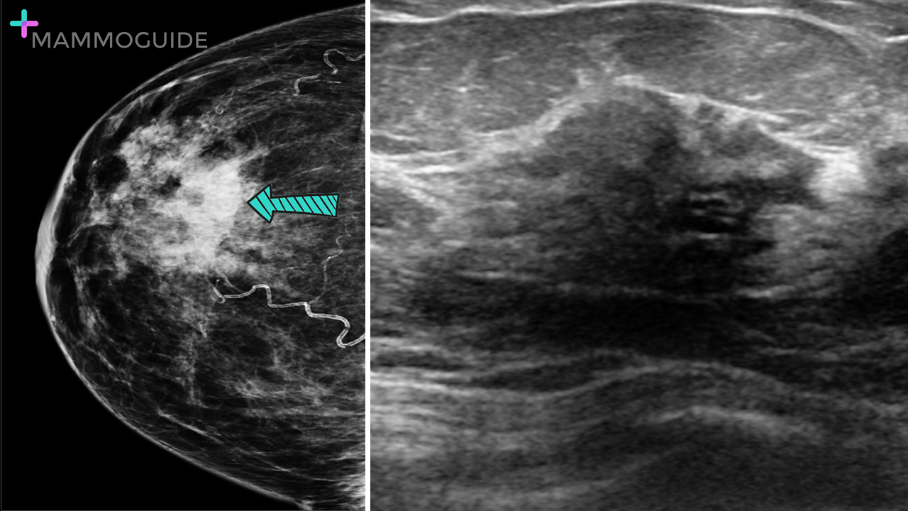 IMAGING FINDINGS:
Craniocaudal view demonstrates an asymmetry in the central right breast. Ultrasound evaluation revealed an irregular, heterogeneous mixed echogenicity mass with ill-defined margins and posterior acoustic shadowing. WHY IT MATTERS:
FURTHER READING: Diabetic Mastopathy: Imaging Features and the Role of Image-guided Biopsy in its Diagnosis (Ultrasonography, 2016)  IMAGING FINDINGS:
Bilateral MLO views demonstrate innumerable high density small round masses within the breasts bilaterally. The patient had a history of direct silicone injections for cosmetic purposes. WHY IT MATTERS:
FURTHER READING: Imaging Spectrum of Extracapsular Silicone: Correlation of US, MR Imaging, Mammographic, and Histopathologic Findings (RadioGraphics, 1999) 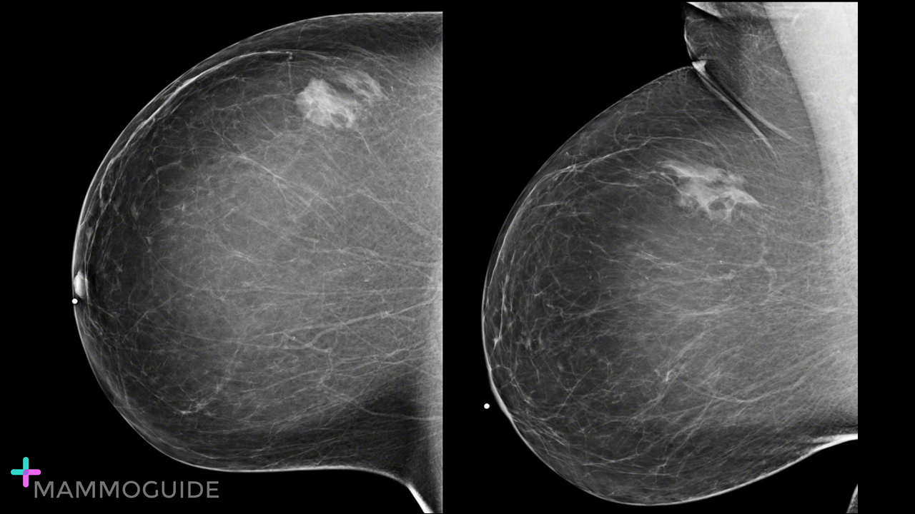 IMAGING FINDINGS:
MLO and CC views of the right breast demonstrate a focal asymmetry in the upper outer quadrant at a posterior depth. WHY IT MATTERS:
|
Quick CasesA picture is worth a thousand words. High-yield examples of essential breast imaging knowledge. 
Categories
All
|
 RSS Feed