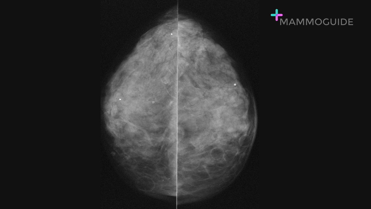
 IMAGING FINDINGS:
Right MLO view demonstrates pleomorphic calcifications in the central right breast in a segmental distribution. WHY IT MATTERS:
FURTHER READING: ACR BI-RADS® Atlas Fifth Edition: Quick Reference
0 Comments
 IMAGING FINDINGS:
Bilateral CC views demonstrate multiple round circumscribed fat density masses within the breasts. Imaging characteristics are consistent with multiple benign oil cysts. WHY IT MATTERS:
FURTHER READING: Steatocystoma Multiplex: Mammographic and Sonographic Manifestations (AJR, 2003) 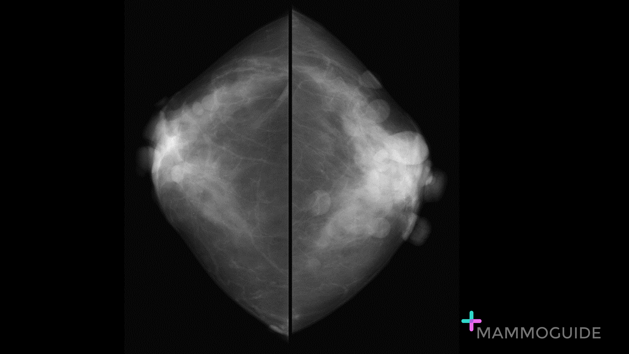 IMAGING FINDINGS:
Bilateral CC views demonstrate multiple superficial round, circumscribed masses bilaterally. WHY IT MATTERS:
FURTHER READING: Increased Risk of Breast Cancer in Neurofibromatosis Type 1: Current Insights (Dove Press, 2017) 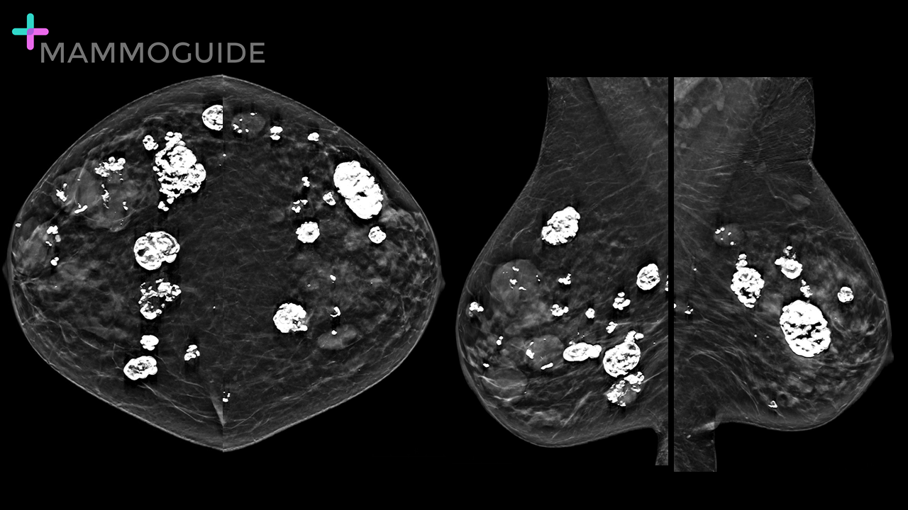 IMAGING FINDINGS:
Bilateral MLO views demonstrate multiple round masses in both breasts. Many of the masses are calcified. WHY IT MATTERS:
FURTHER READING: Multiple Bilateral Masses Detected on Screening Mammography: Assessment of Need for Recall Imaging (AJR, 2000) 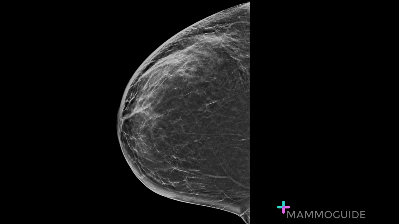 IMAGING FINDINGS:
Craniocaudal view of the right breast demonstrates diffuse skin thickening. There is no underlying mass visualized. WHY IT MATTERS:
FURTHER READING: Breast Emergencies: Types, Imaging Features, and Management (AJR) 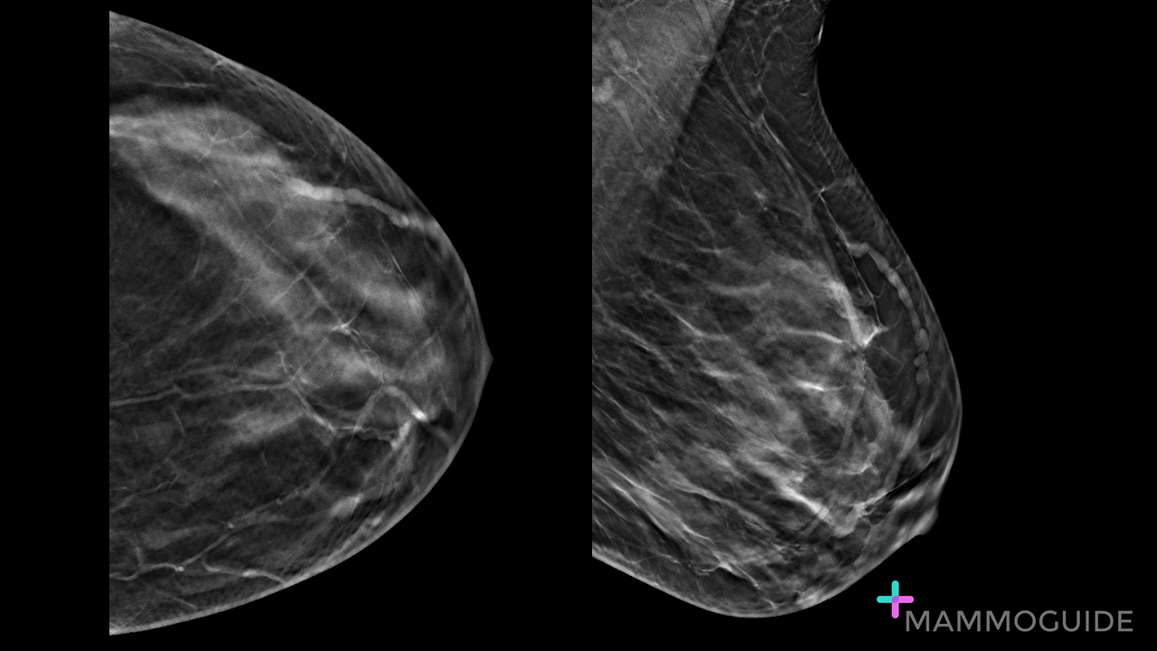 IMAGING FINDINGS:
CC and MLO tomosynthesis views of the left breast show a dilated tubular structure superficially in the upper outer quadrant. WHY IT MATTERS:
Mondor’s Disease of the Breast: Sonographic and Mammographic Findings (AJR, 2000) 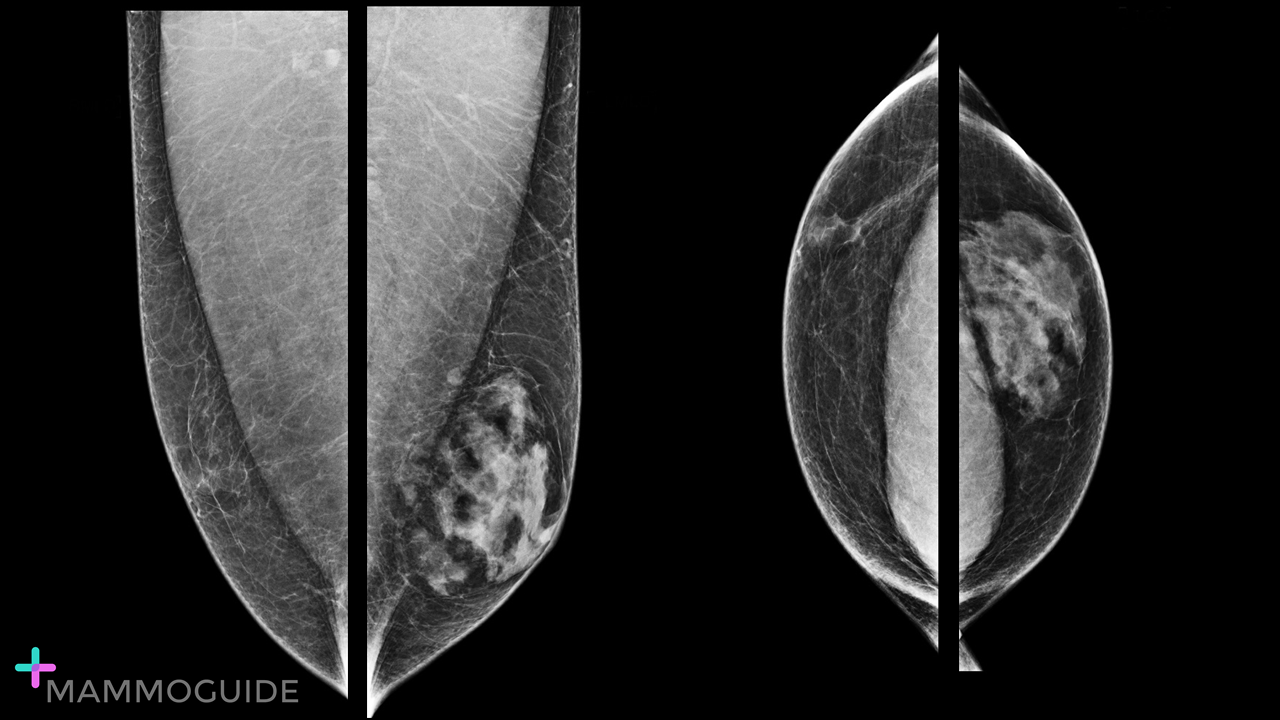 IMAGING FINDINGS:
Mammographic views demonstrate a normal male tissue pattern in the right breast and moderate to marked gynecomastia in the left breast. WHY IT MATTERS:
FURTHER READING: Illuminations: Three Patterns of Male Gynecomastia (RadioGraphics, 2013) 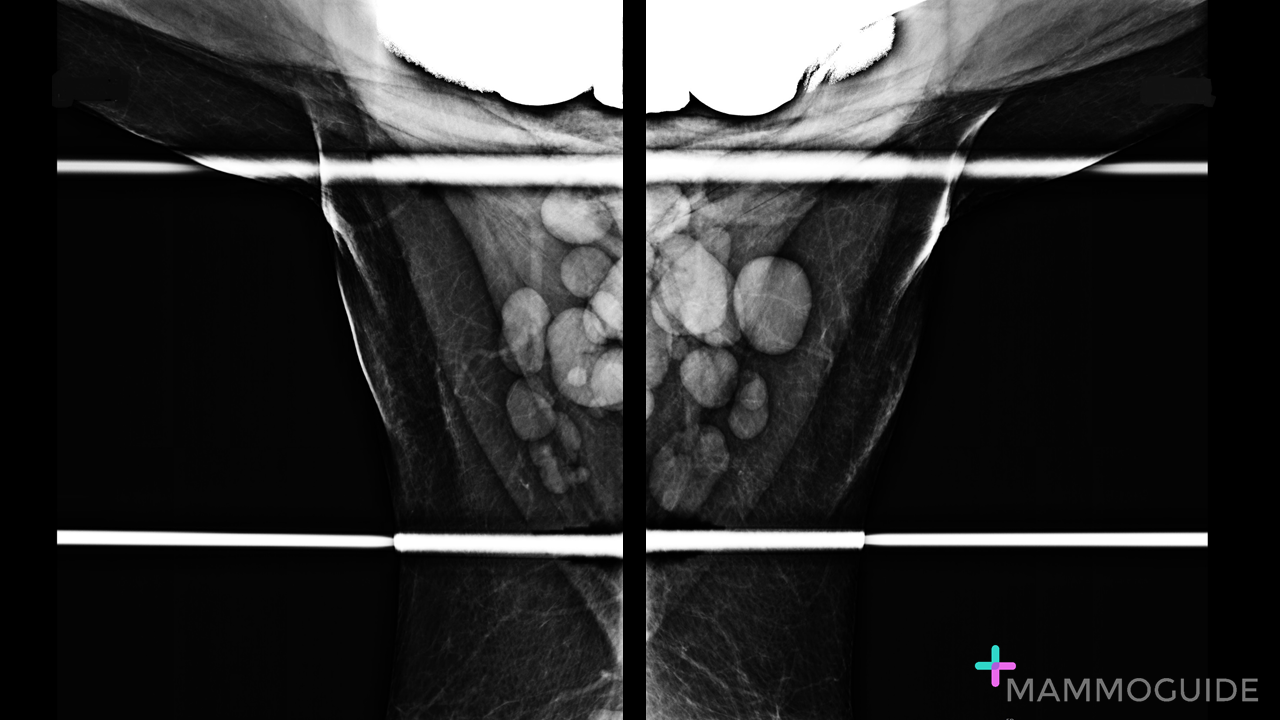 IMAGING FINDINGS:
Axillary views demonstrate diffuse, symmetric bilateral axillary lymphadenopathy. WHY IT MATTERS:
FURTHER READING: Mammographic Signs of Systemic Disease (RadioGraphics, 2011) 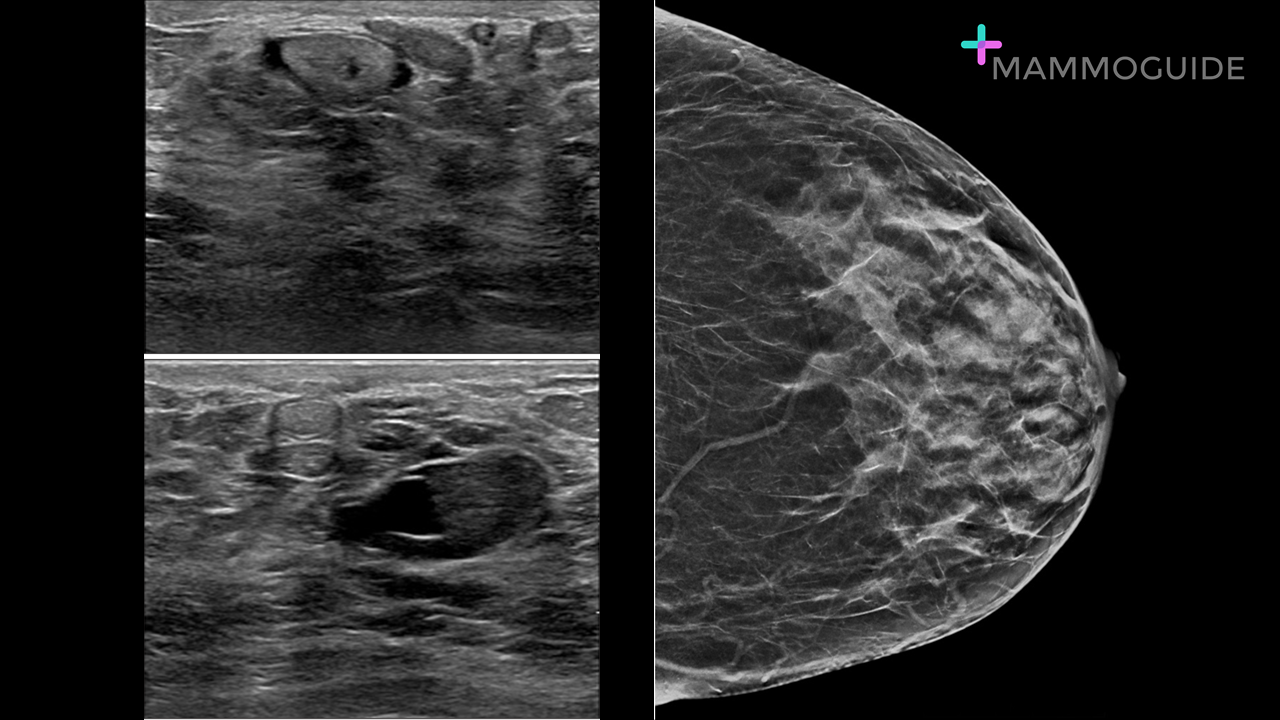 IMAGING FINDINGS:
Breast ultrasound demonstrates anechoic, tubular branching ducts in the subareolar region. There is echogenic debris present within the ducts. Mammography demonstrates dilated ducts and mixed density masses in the subareolar region. WHY IT MATTERS:
FURTHER READING: Imaging Approaches to Diagnosis and Management of Common Ductal Abnormalities (RadioGraphics, 2012) 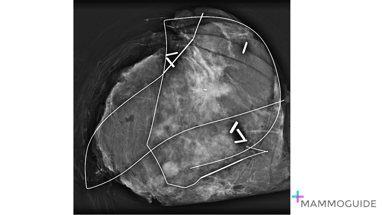 IMAGING FINDINGS:
Specimen image shows a bracketed wire localization of a spiculated mass with associated pleomorphic calcifications. WHY IT MATTERS:
What to look for on a Breast Specimen Radiograph: Lessons Learnt (BMJ Case Rep, 2015) |
Quick CasesA picture is worth a thousand words. High-yield examples of essential breast imaging knowledge. 
Categories
All
|
 RSS Feed