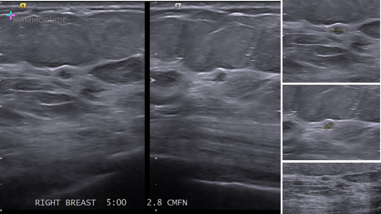
 IMAGING FINDINGS:
Multiple sonographic images obtained at the 5:00 position of the right breast demonstrate normal fibroglandular tissue. A “lesion” was identified and measured in two planes. Color images and harmonic images were also performed for this 1 – 2 mm “lesion.” This case then went on to another institution for a second opinion. WHY IT MATTERS:
FURTHER READING: Breast Ultrasonography: State of the Art (RSNA 2013)
0 Comments
Leave a Reply. |
Quick CasesA picture is worth a thousand words. High-yield examples of essential breast imaging knowledge. 
Categories
All
|
 RSS Feed