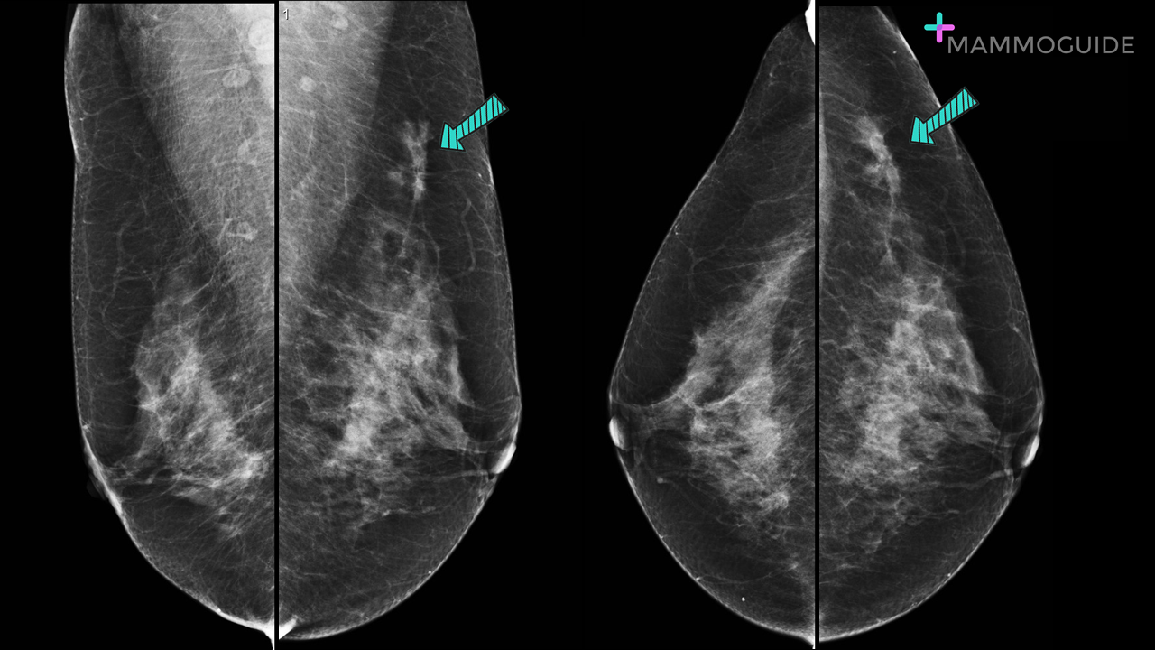
 IMAGING FINDINGS:
Standard craniocaudal and mediolateral oblique views of both breasts demonstrate a focal asymmetry in the upper outer left breast. There is suggestion of associated architectural distortion. WHY IT MATTERS:
Focal Asymmetric Densities Seen at Mammography: US and Pathologic Correlation (RadioGraphics 2002)
0 Comments
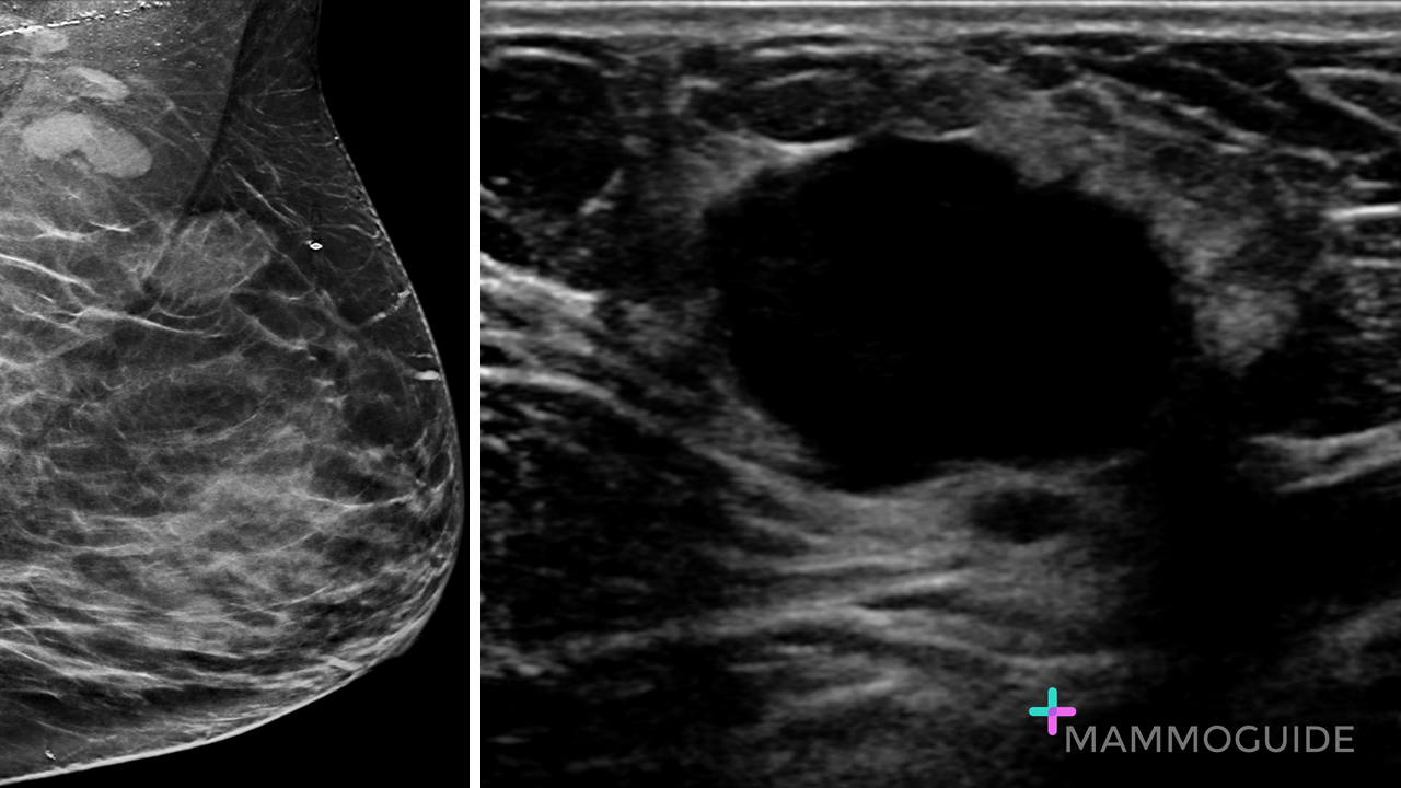 IMAGING FINDINGS:
A mediolateral oblique view demonstrates a large mass with obscured margins in the superior left breast. There are also enlarged left axillary lymph nodes. An ultrasound image demonstrates a large “anechoic” mass. This was said to be a “cyst” by the sonographer. However, the gain is incorrect on this image. With appropriate sonographic settings this proved to be a suspicious hypoechoic mass and ultimately an invasive ductal carcinoma (which is what was expected based on the mammographic findings.) WHY IT MATTERS:
FURTHER READING: Breast Ultrasonography: State of the Art (Radiology 2013) 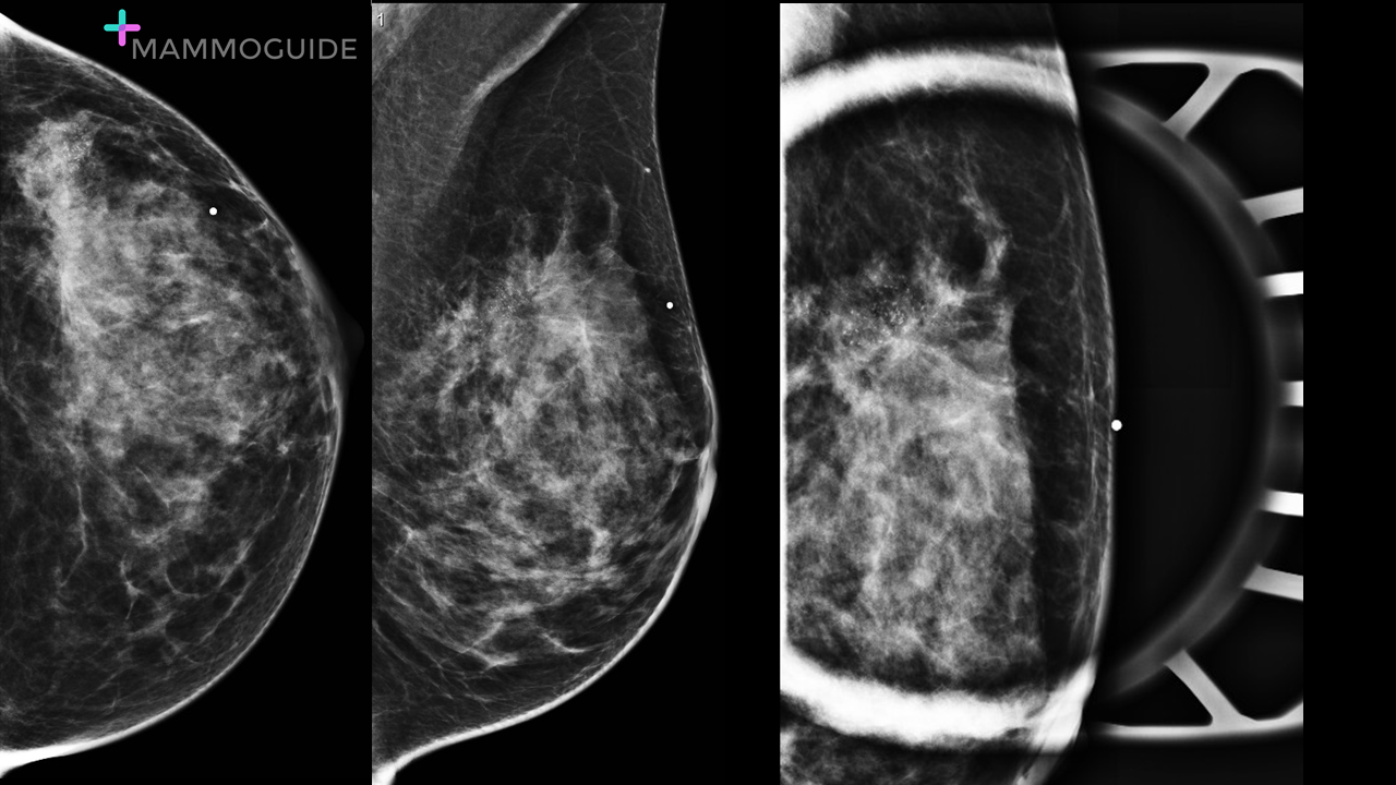 IMAGING FINDINGS:
Craniocaudal and mediolateral oblique views of the left breast demonstrate a suspicious spiculated mass with associated pleomorphic microcalcifications. A BB is placed on the skin to indicate that the mass is palpable. A tangential magnified view of the palpable mass again demonstrates the suspicious spiculated mass with calcifications in the area of clinical concern. WHY IT MATTERS:
FURTHER READING: Imaging Management of Palpable Breast Abnormalities 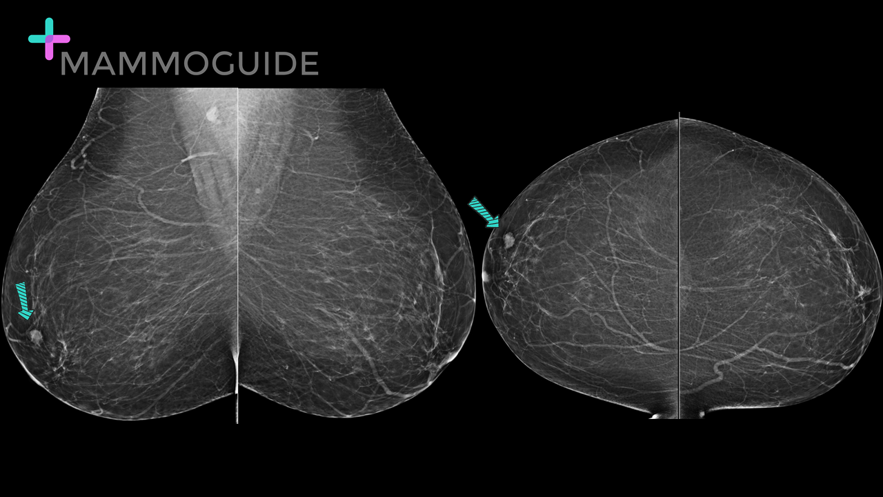 IMAGING FINDINGS:
Standard mediolateral oblique (MLO) and craniocaudal (CC) views of a patient with breasts composed almost entirely of fat demonstrate an oval hyperdense mass with irregular margins in the subareolar right breast. WHY IT MATTERS:
FURTHER READING: Nipple-Areolar Complex: Normal Anatomy and Benign and Malignant Processes 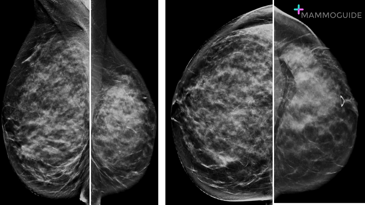 IMAGING FINDINGS:
Bilateral CC and MLO views demonstrate dramatic asymmetry in the size of the breasts. The left breast is significantly smaller than the right breast. This is a change compared to prior exams. WHY IT MATTERS:
FURTHER READING: The Shrinking Breast: An Unusual Mammographic Finding of Invasive Lobular Carcinoma (Radiology Case Reports 2007) 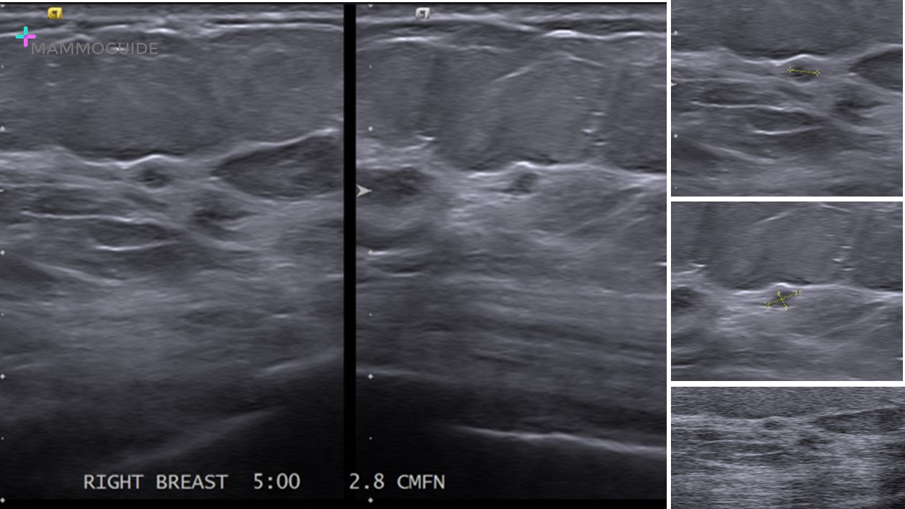 IMAGING FINDINGS:
Multiple sonographic images obtained at the 5:00 position of the right breast demonstrate normal fibroglandular tissue. A “lesion” was identified and measured in two planes. Color images and harmonic images were also performed for this 1 – 2 mm “lesion.” This case then went on to another institution for a second opinion. WHY IT MATTERS:
FURTHER READING: Breast Ultrasonography: State of the Art (RSNA 2013) 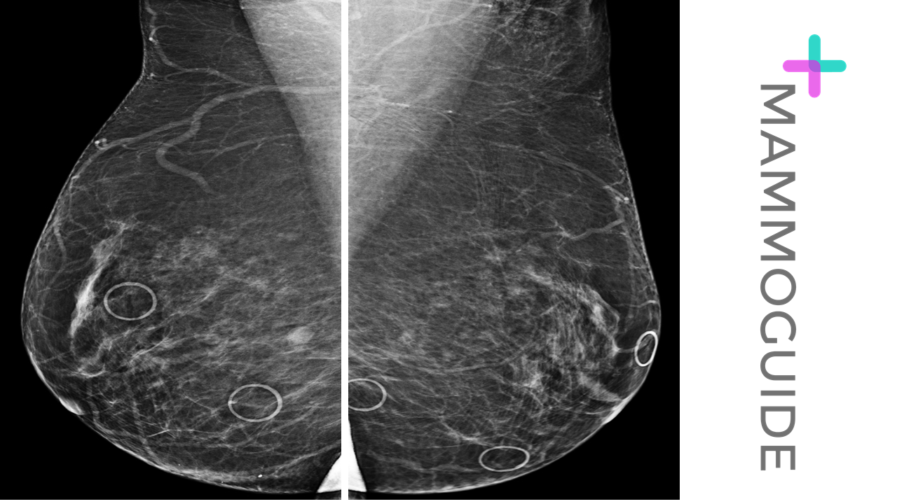 IMAGING FINDINGS:
Bilateral mediolateral oblique (MLO) views demonstrate breasts composed of scattered fibroglandular elements with no apparent abnormality. Multiple mole markers are placed on the breasts for the exam, which is an unnecessary step. WHY IT MATTERS:
Digital Breast Tomosynthesis: Lessons Learned from Early Clinical Implementation (RadioGraphics 2014) 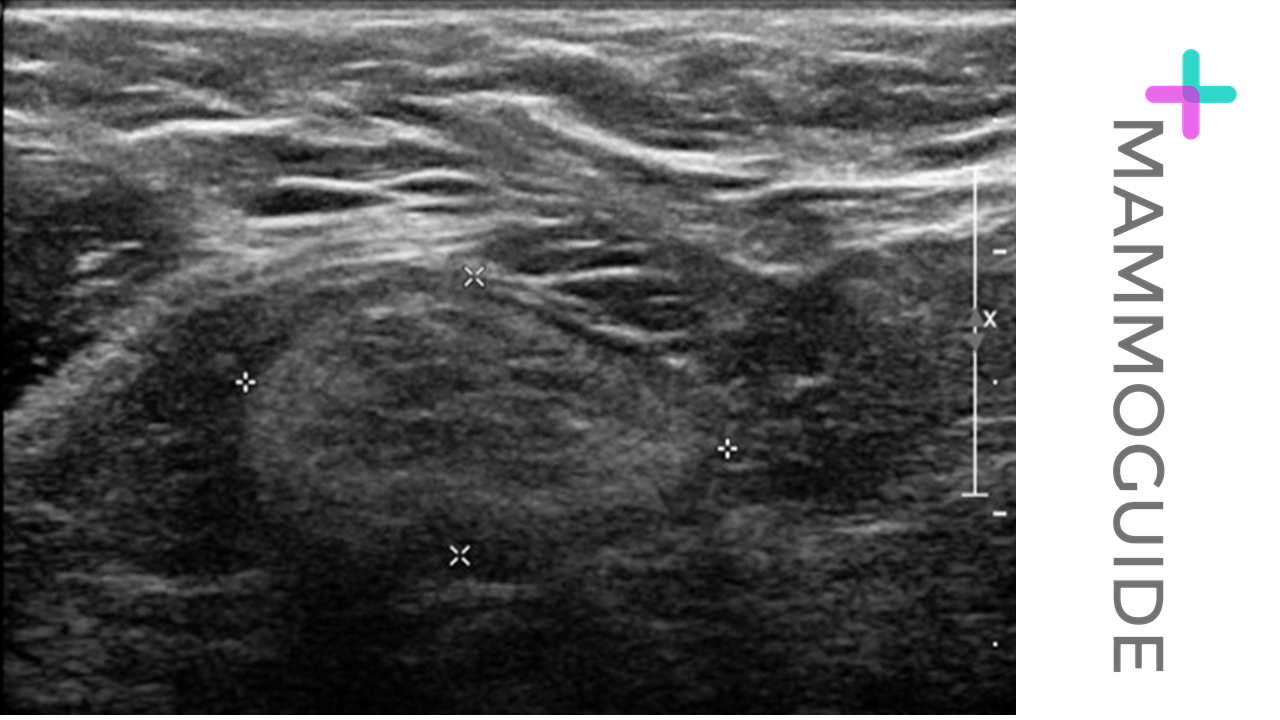 IMAGING FINDINGS:
Sonographic image of the right axilla demonstrates a normal axillary lymph node with a thin cortex. The sonographer (or radiologist) is incorrectly measuring the dimensions of the lymph node. WHY IT MATTERS:
FURTHER READING: Axillary Staging of Breast Cancer: What the Radiologist Should Know (RadioGraphics 2013) 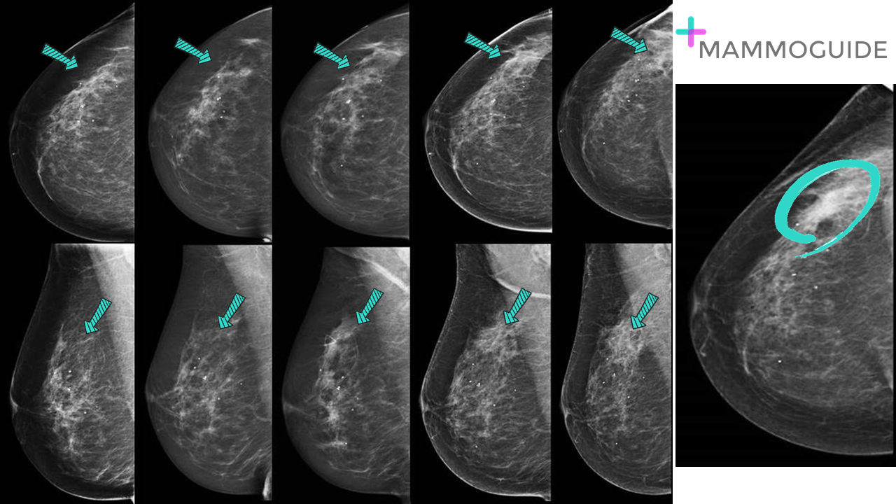 IMAGING FINDINGS:
Serial mammograms of the right breast over five years. There is a developing asymmetry in the right breast. Notice the gradual increase in density in the upper outer right breast without an obvious mass. It is difficult to appreciate an abnormality on any given year, however comparison with multiple prior studies reveals a definite change. WHY IT MATTERS:
FURTHER READING: The Developing Asymmetry: Revisiting a Perceptual and Diagnostic Challenge (RSNA 2015) 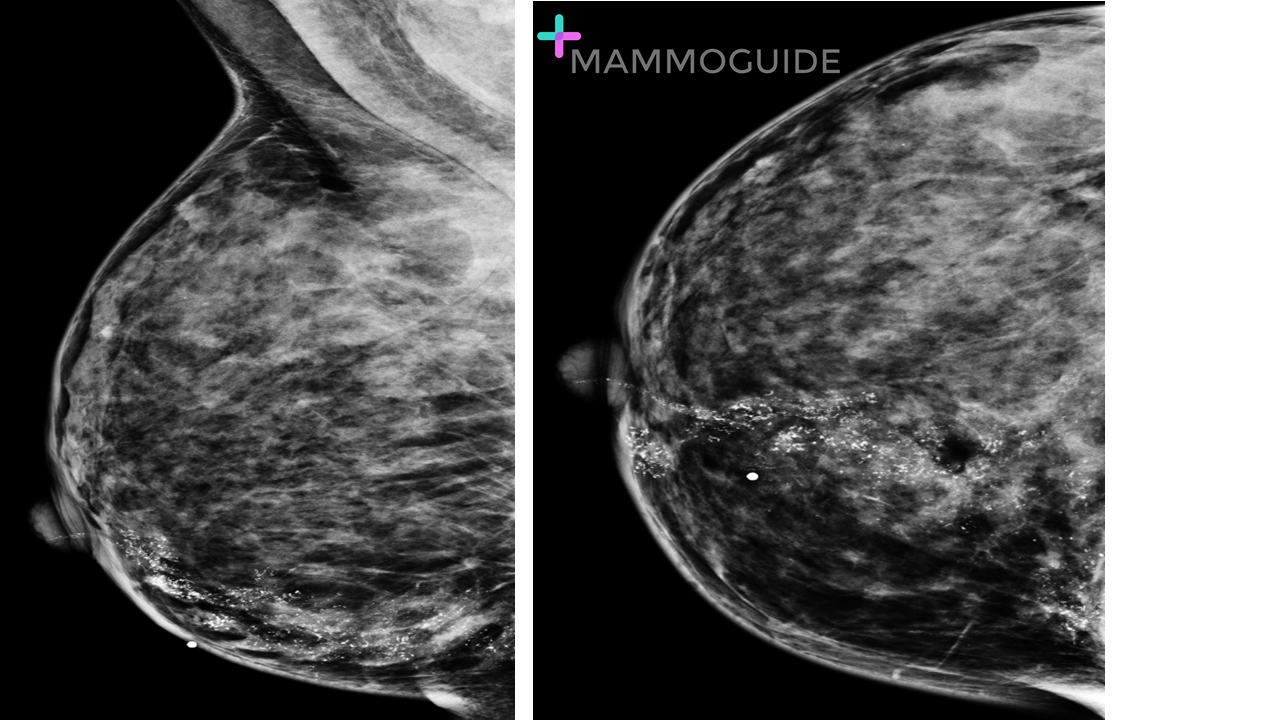 IMAGING FINDINGS:
Standard mediolateral oblique (MLO) and Craniocaudal (CC) views of the right breast demonstrate extensive pleomorphic calcifications in a segmental distribution extending from the posterior chest wall to the nipple. WHY IT MATTERS:
FURTHER READING: High‐Grade Ductal Carcinoma In Situ: An Overview for the Radiologist (J Am Osteopath Coll Radiol 2013) |
Quick CasesA picture is worth a thousand words. High-yield examples of essential breast imaging knowledge. 
Categories
All
|
 RSS Feed