 IMAGING FINDINGS:
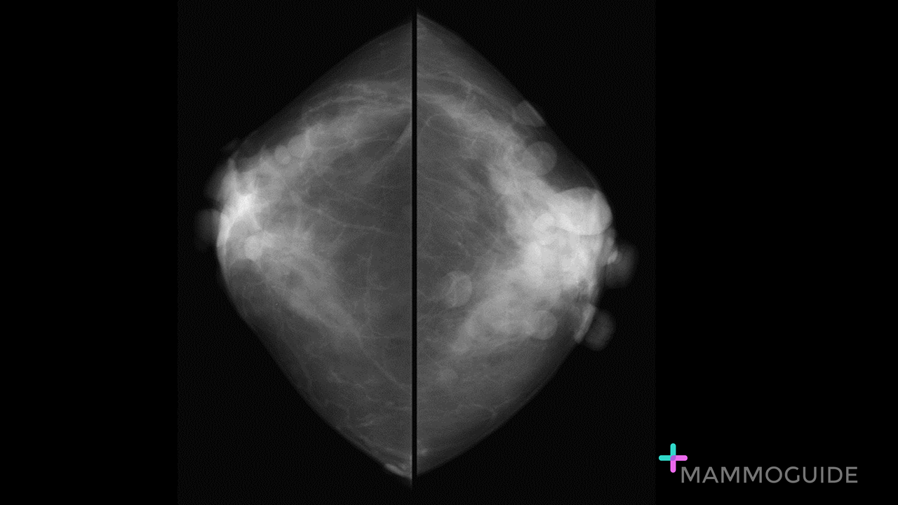
Bilateral CC views demonstrate multiple superficial round, circumscribed masses bilaterally. WHY IT MATTERS:
FURTHER READING: Increased Risk of Breast Cancer in Neurofibromatosis Type 1: Current Insights (Dove Press, 2017)
0 Comments
 IMAGING FINDINGS:
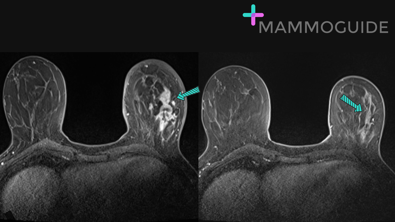
The first post-contrast axial MRI image shows segmental nonmass enhancement involving the majority of the lateral left breast. The second post-contrast axial image, performed after 6 rounds of neo-adjuvant chemotherapy, demonstrates complete imaging response to therapy with decrease in size of the lesion and no remaining enhancement identified. WHY IT MATTERS:
Imaging Neoadjuvant Therapy Response in Breast Cancer (RSNA, 2017) 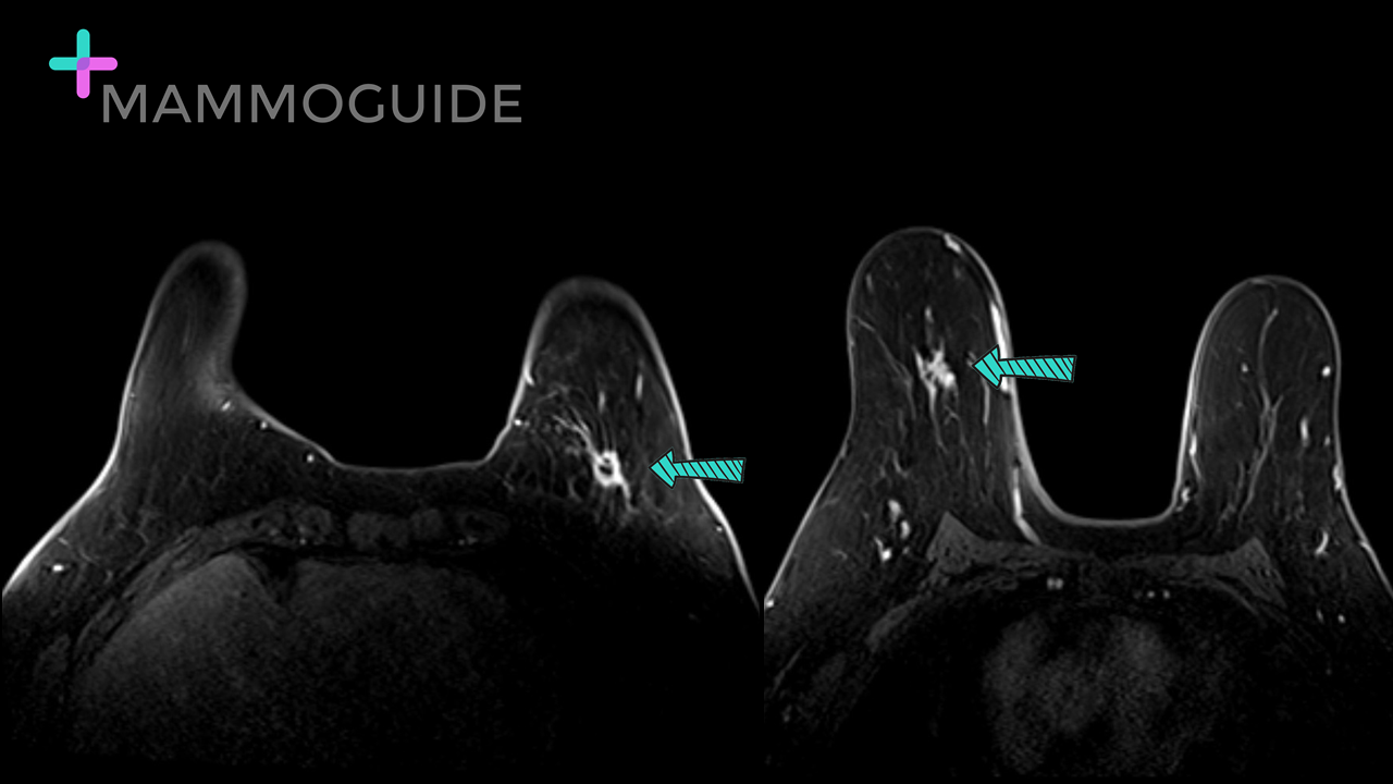 IMAGING FINDINGS:
Post-contrast breast MRI image performed to assess extent of disease demonstrate the known biopsy proven malignancy (index lesion) in the left breast. There is a rim enhancing mass containing a biopsy clip in the inferior left breast at a posterior depth. The second post-contrast image demonstrates an irregular, enhancing mass in the central right breast at a middle depth. This is highly suspicious for contralateral disease. WHY IT MATTERS:
FURTHER READING: MRI Evaluation of the Contralateral Breast in Women with Recently Diagnosed Breast Cancer (N Engl J Med, 2007) 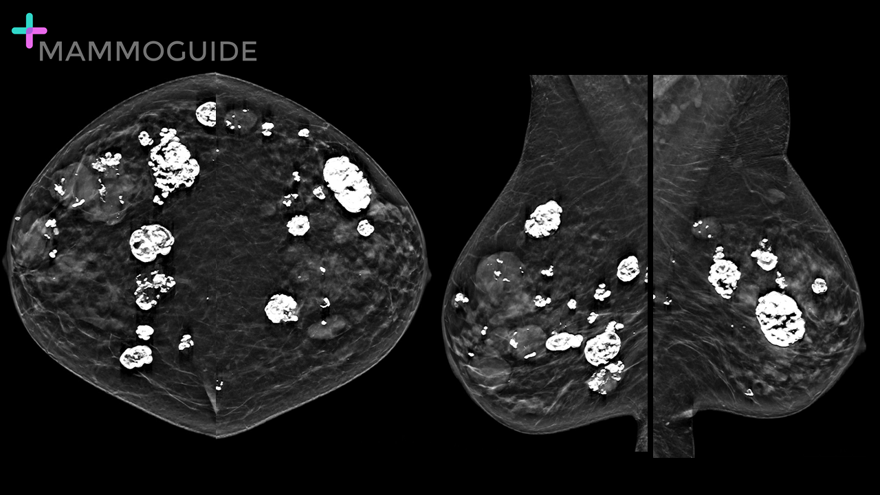 IMAGING FINDINGS:
Bilateral MLO views demonstrate multiple round masses in both breasts. Many of the masses are calcified. WHY IT MATTERS:
FURTHER READING: Multiple Bilateral Masses Detected on Screening Mammography: Assessment of Need for Recall Imaging (AJR, 2000) 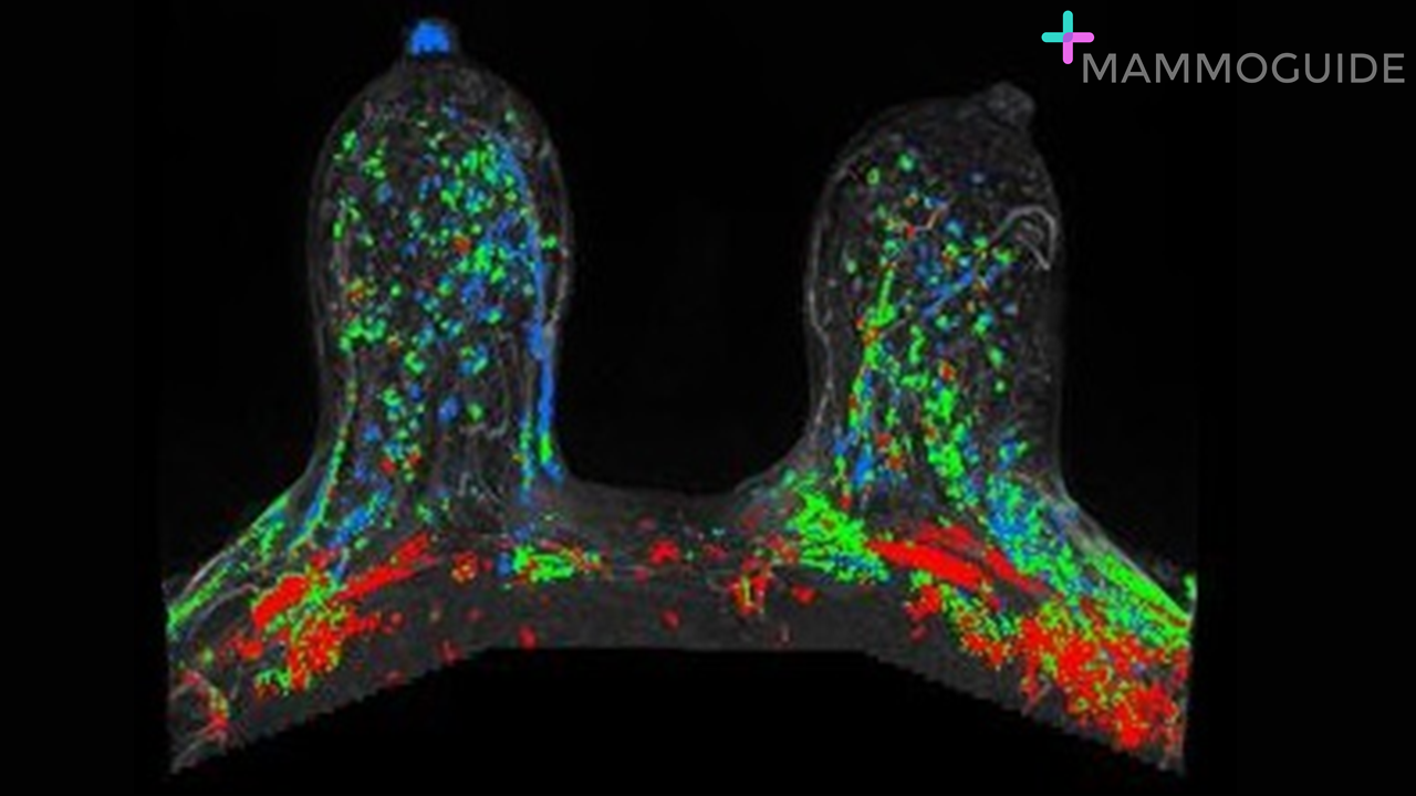 IMAGING FINDINGS:
Maximum intensity projection (MIP) image demonstrates moderate to marked symmetric background parenchymal enhancement bilaterally. WHY IT MATTERS: 1. Background parenchymal enhancement is reported on all breast MRI’s. The four categories are: a. Minimal b. Mild c. Moderate d. Marked 2. Background parenchymal enhancement is assessed as either symmetric or asymmetric. The MIP image gives a good overview of both breasts. FURTHER READING: Background Parenchymal Enhancement at Breast MR Imaging: Normal Patterns, Diagnostic Challenges, and Potential for False-Positive and False-Negative Interpretation (RadioGraphics, 2014) 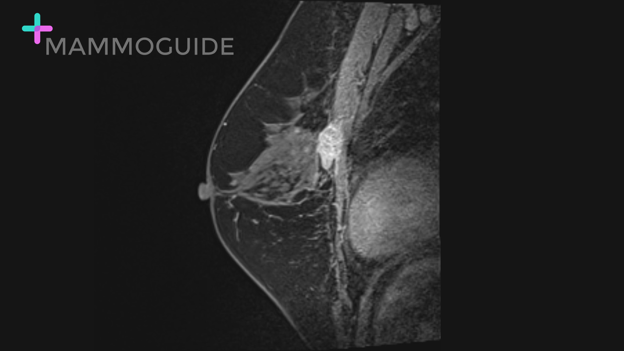 IMAGING FINDINGS:
Sagittal MRI image demonstrates an irregular enhancing mass in the inferior right breast at a posterior depth. There is enhancement of the underlying pectoralis muscle consistent with pectoralis invasion. WHY IT MATTERS:
FURTHER READING: Radiologists’ Role in Breast Cancer Staging: Providing Key Information for Clinicians (RadioGraphics, 2014) 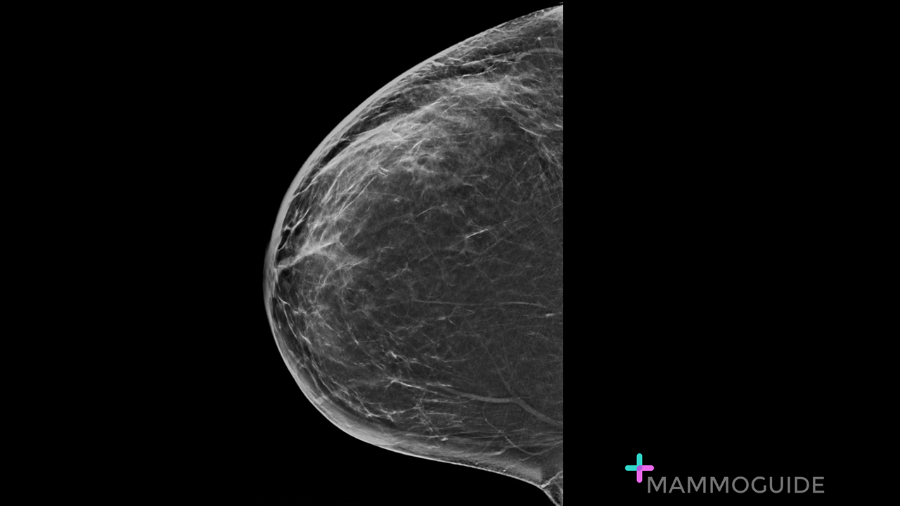 IMAGING FINDINGS:
Craniocaudal view of the right breast demonstrates diffuse skin thickening. There is no underlying mass visualized. WHY IT MATTERS:
FURTHER READING: Breast Emergencies: Types, Imaging Features, and Management (AJR) 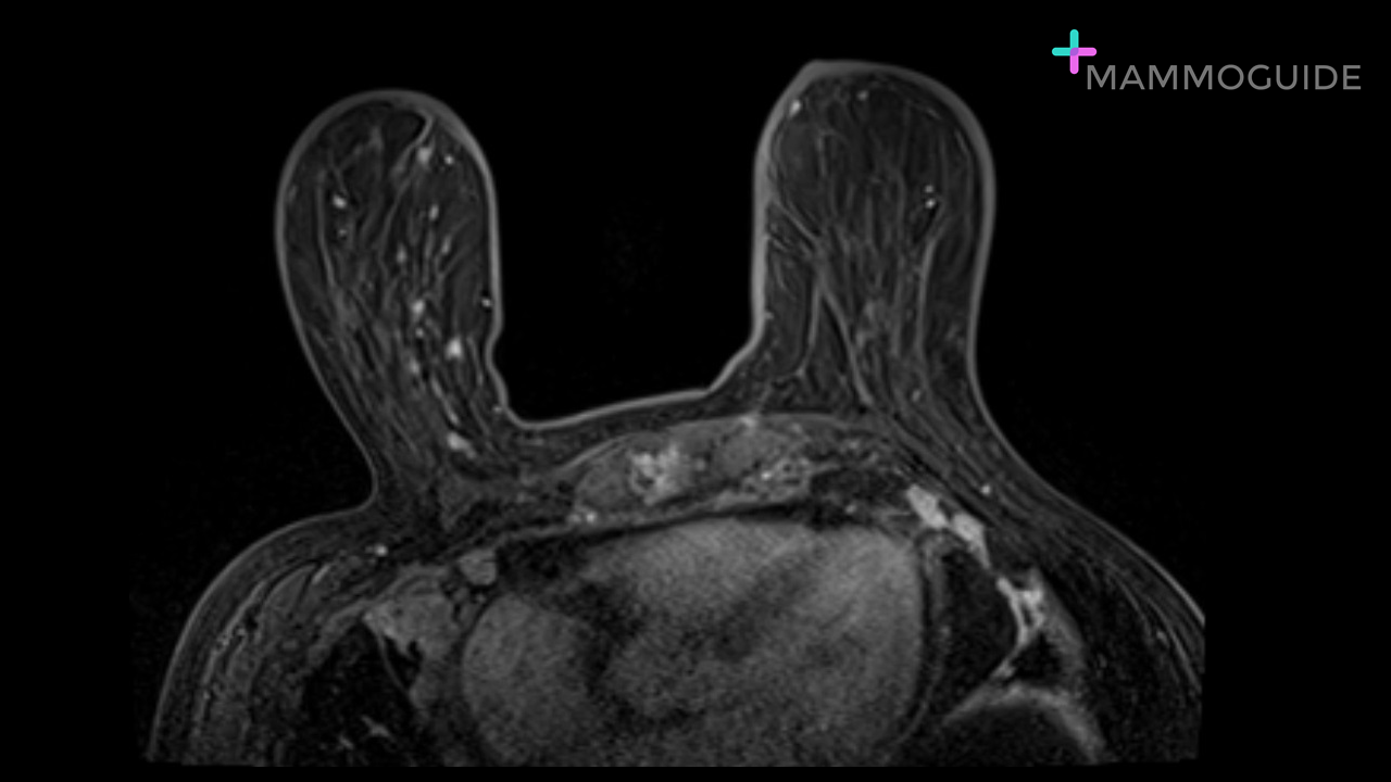 IMAGING FINDINGS:
Axial post-contrast image demonstrates a heterogeneously enhancing mass involving the sternum. There are also enhancing lesions in several ribs. WHY IT MATTERS:
FURTHER READING: Delineating Extramammary Findings at Breast MR Imaging (RadioGraphics, 2017) 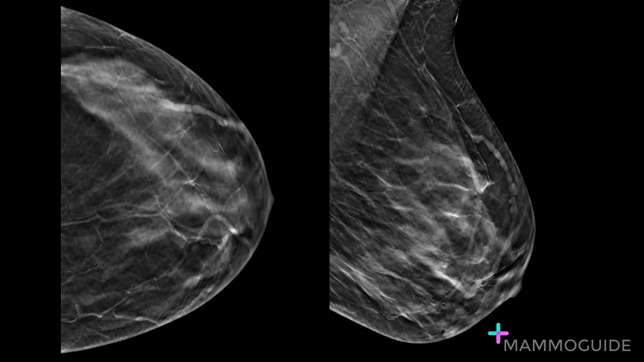 IMAGING FINDINGS:
CC and MLO tomosynthesis views of the left breast show a dilated tubular structure superficially in the upper outer quadrant. WHY IT MATTERS:
Mondor’s Disease of the Breast: Sonographic and Mammographic Findings (AJR, 2000) 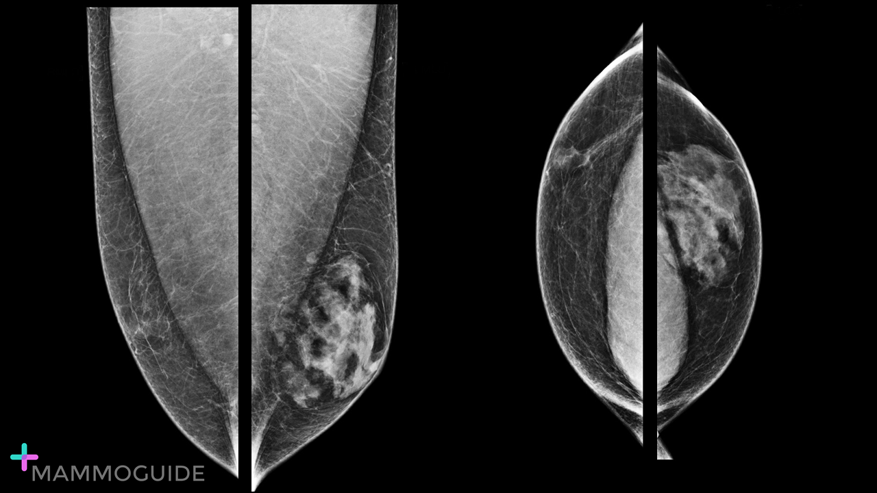 IMAGING FINDINGS:
Mammographic views demonstrate a normal male tissue pattern in the right breast and moderate to marked gynecomastia in the left breast. WHY IT MATTERS:
FURTHER READING: Illuminations: Three Patterns of Male Gynecomastia (RadioGraphics, 2013) |
Quick CasesA picture is worth a thousand words. High-yield examples of essential breast imaging knowledge. 
Categories
All
|
 RSS Feed